Bachstetter, Spinal Cord and Brain Injury Research Center, Department of Neuroscience, University of Kentucky, 741 S. Limestone Street, BBSRB Room B459, Lexington, Kentucky 40536–0509, USA. Phone: 859.218.4315; Email: [email protected].
Bachstetter, Spinal Cord and Brain Injury Research Center, Department of Neuroscience, University of Kentucky, 741 S. Limestone Street, BBSRB Room B459, Lexington, Kentucky 40536–0509, USA. Phone: 859.218.4315; Email: [email protected].
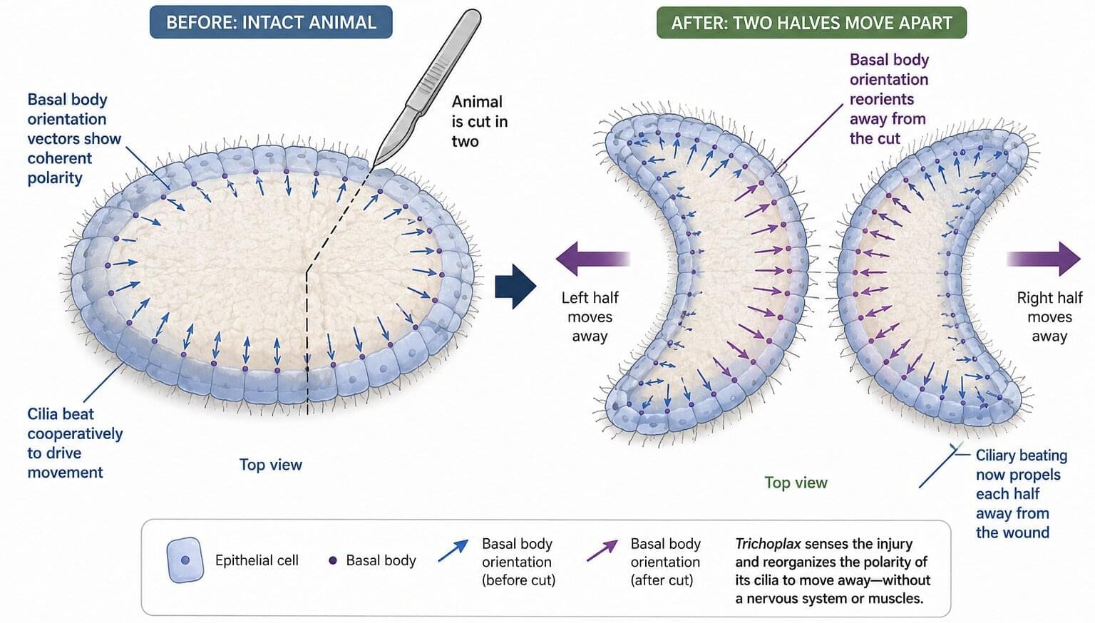
For a flat sea creature just a few millimeters across, a gentle poke is instantly recognized as danger. Trichoplax adhaerens—a translucent blob with no head, brain or muscles—scuttles away in seconds when touched. Imagine a flattened multicellular amoeba moving as a single unit: Trichoplax is only ~20 microns thick and a few millimeters wide. It glides on surfaces by beating tens of thousands of cilia on its lower epithelium (the underside), like microscopic oars dragging against the water.
Yet unlike most animals, Trichoplax has no obvious front or back end, no nerves or muscles at all. How can such a simple “crawling carpet” steer or change direction without a brain?
A new study reveals the remarkable flexibility of this pinhead-sized animal. While in most creatures, the orientation of each cilium is fixed early in development and locked to the body’s axes, Trichoplax achieves its swift escape by reorienting its thousands of hairlike cilia.
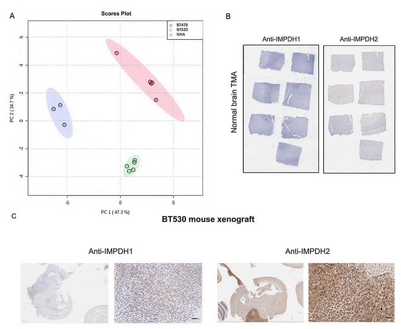
A new study has identified a more precise and effective way to prevent cancer from spreading to the brain. The paper, published in the Proceedings of the National Academy of Sciences, details the development of novel drug candidates that target a key enzyme implicated in the spread of lung, breast, skin and other cancers to the brain. The work builds on a promising new therapeutic strategy first reported by the same group of researchers last year.
The new drug candidates are designed to intercept rogue cancer cells before they depart from primary tumors and ultimately travel to the brain.
Lead author Sheila Singh, based at both King’s College London and McMaster University, says this type of cancer—called metastatic brain cancer—is the most common type of brain tumor in adults and comes with an extremely grim outlook, with 90% of patients dying within one year of diagnosis.
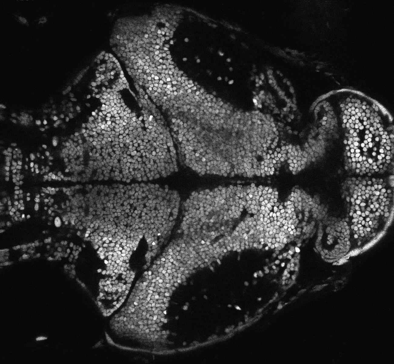
The altered presence of tiny fragments of neuronal genes, called microexons, causes hyperarousal in zebrafish. This is the main conclusion of an international study led by Pompeu Fabra University (UPF) and the Center for Genomic Regulation (CRG). An abnormal pattern of neural microexon presence leads to a hyperarousal state characterized by heightened neural activity and insomnia, commonly associated with stress but also with neurodevelopmental disorders.
Arousal regulation is highly conserved in evolution. Therefore, this finding could help researchers understand the mechanism underlying some human neurodevelopmental disorders, such as autism and schizophrenia, conditions associated with microexon mutations.
To survive, animals need to be ready to react to external and internal stimuli. This activation of the central nervous system, arousal, is highly conserved throughout the animal kingdom.
Microplastics – tiny bits of degraded polymers that are ubiquitous in our air, water and soil – have lodged themselves throughout the human body, including the liver, kidney, placenta and testes, over the past half century.
Now, University of New Mexico Health Sciences researchers have found microplastics in human brains, and at much higher concentrations than in other organs. Worse, the plastic accumulation appears to be growing over time, having increased by 50% over just the past eight years.
In a new study published in Nature Medicine, a team led by toxicologist Matthew Campen, PhD, Distinguished and Regents’ Professor in the UNM College of Pharmacy, reported that plastic concentrations in the brain appeared higher than in the liver or kidney, and higher than previous reports for placentas and testes.
Aeran and colleagues present research on targeted gene therapy vector engineering and pre-clinical testing of neuron-targeted AAV9-based constructs for STXBP1-related neurodevelopmental and epileptic encephalopathies. Candidate vectors designed to target specific neuronal types and detarget tissues associated with toxicity produced robust phenotypic reversal in Stxbp1 +/− mice and were well tolerated in monkeys.
In this Presidential Lecture, cognitive scientist Douglas Hofstadter examines the role and contributions of analogy in cognition, using a variety of analogies to illustrate his points.
Stanford University:
Stanford Humanities Center:
Stanford University Channel on YouTube:
I think this was one of my most enjoyable dialogues in our What’s new series. Maybe Sabine and I are getting more used to each other’s cadence and interests or maybe it was the subject matter. Either way, I think you will find this to be a fascinating and provocative discussion of science at the forefront, and at the not-so-forefront, because that science is interesting too! We began our discussion describing a new finding of a Giant Ring of galaxies billions of light years across in the sky. The key questions are: Is it real? And is it surprising? We both have slightly different takes on this. Next we described a new measurement of the strength of gravity on scales from 80 to 800 million light years in distance. And guess what? Gravity falls off just like Newton predicted! This may seem like a big yawn, but one of the most popular models that claims to do away with dark matter would imply that Gravity would fall off differently on these scales. Does this new result kill that idea? Stay tuned. Microsoft, which has cried wolf a number of times so far when it comes to something called Majorana qubits as the basis of a new viable quantum computer just published a new paper claiming they finally have it. Sabine and I discuss why we are both still skeptical, but why the effort is worth it. Next, CERN, the large European particle physics laboratory, and the world particle physics community seem to have converged on plans for building a huge new accelerator in the current CERN site… this time involving an underground ring 91 km in circumference, in which electrons and positrons would collide to explore the detailed properties of the Higgs particle. Is the effort worth it? Again, Sabine and I have slightly different takes on this. Fusion power, which we have talked about in a number of earlier episodes, continues to tempt humanity with the promise of unlimited energy. Many people, myself included, have tended to argue that fusion seems to be 25 years in the future, and may always be 25 years in the future. But many new efforts are underway, so who knows. Unfortunately, a group of economists has analyzed fusion in the context of other large energy programs and have argued that even if we can achieve it, it may not be as economically viable as many claim. Finally, one day Richard Feynman went to a Thai restaurant with his young companion Ralph Leighton, and wondered what he should order. Should it be the same old dish he loved or something new. An equation filled napkin later, and he had the answer. Fifty years later some cognitive scientists resurrected Feynman’s napkin and explained it, and argued it might have important implications in other social situations. Such is the power of science. Consider supporting the podcast and the Origins Project Foundation at https://www.originsproject.org/ To see commercial-free, full HD video episodes, join us at lawrence krauss.substack.com Thank you for your support! iTunes: https://podcasts.apple.com/us/podcast… https://TheOriginsPodcast.com Twitter: / theoriginspod Instagram:
/ theoriginspod Facebook:
/ theoriginspod The Origins Podcast, a production of The Origins Project Foundation, features in-depth conversations with some of the most interesting people in the world about the issues that impact all of us in the 21st century. Host, theoretical physicist, lecturer, and author, Lawrence M. Krauss, will be joined by guests from a wide range of fields, including science, the arts, and journalism. The topics discussed on The Origins Podcast reflect the full range of the human experience — exploring science and culture in a way that seeks to entertain, educate, and inspire. Full Episodes Playlist:
• Ricky Gervais — The Origins Podcast with L…
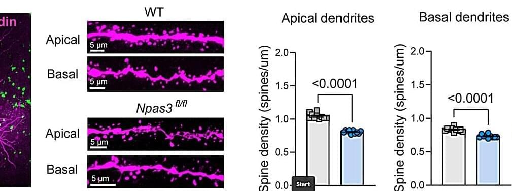
Researchers in the Jacobs School of Medicine and Biomedical Sciences at the University at Buffalo have discovered a connection between a specific gene and healthy brain function. “The hope is that this discovery could eventually lead to expanded treatment for psychiatric and neurological disorders such as schizophrenia, bipolar disorder and autism,” explains Mikhail V. Pletnikov, MD, Ph.D., professor and chair of the Department of Physiology and Biophysics, the senior author of the study with Kateryna (Kate) Murlanova, Ph.D., the first lead author and a research scientist in the department.
They discovered that the NPAS3 gene expressed in astrocytes—the cells that help with brain chemistry—regulates the energy production required to support thinking and memory. NPAS3 is a transcription factor, which means it directs how certain genes work and influences how cells function. Their findings are published in Science Advances.
“Previous studies have linked NPAS3 to conditions involving cognitive problems, such as schizophrenia, but scientists didn’t know exactly how it might be involved,” Pletnikov says.