Evidence is mounting that there are distinct subtypes of autism, and now, scientists have found that the condition can vary according to the strength of people’s brain connections
A massive new meta-analysis reveals that individual cognitive abilities, like reading and math, rely on inherited DNA just as much as overall intelligence, suggesting people possess heavily customized genetic cognitive profiles independent of general smarts.
Elon Musk UPDATE Neuralink 4.0 Chip introduces Neuralink’s next-generation O1 brain chip developed with Samsung.
This video explores the latest progress of the Neuralink 4.0 chip, including movement restoration, speech recovery, Blindsight vision technology, and how Neuralink patients are using brain-computer interfaces today.
We also examine Samsung’s 4nm partnership, the new R1 surgical robot, and competition from Synchron, Paradromics, and China’s NEO system to understand how the Neuralink 4.0 chip could shape the future of the BCI industry.
If you’re interested in Elon Musk, AI, neuroscience, and future medical technology, this breakdown explains why many experts view the Neuralink 4.0 chip as one of the most important developments in brain-computer interfaces.
🔔 Join our community and hit Subscribe!
https://bit.ly/3i7gILj.
===
#teslacarworld.
#Neuralink.
#Neuralinkupdate.
#BrainComputerInterface.
#Neuralink40Chip.
#BCITechnology

Gary Marcus is now one of the loudest skeptics of the AI boom. In 2012, almost nobody was listening.
I have the tape.
That year, I sat down with him for Singularity. FM, right after he published a sharp critique of Ray Kurzweil’s theory of mind in The New Yorker. Marcus was already making the argument that would define his career. Intelligence is not just pattern-matching. The mind is a kluge, a messy evolutionary patch job. And scale alone will not get you to real #AI.
More than a decade later, that argument is everywhere. Labs are chasing the hybrid and neurosymbolic approaches he pointed to back then. The field finally caught up to the conversation.
But here is what makes the interview worth revisiting. He also bet big on neuroscience as the road forward, on projects like Blue Brain and Whole Brain Emulation. The breakthroughs came from somewhere else entirely.
So was he the prophet, or just early on some calls and wrong on others? Watch it and decide for yourself.
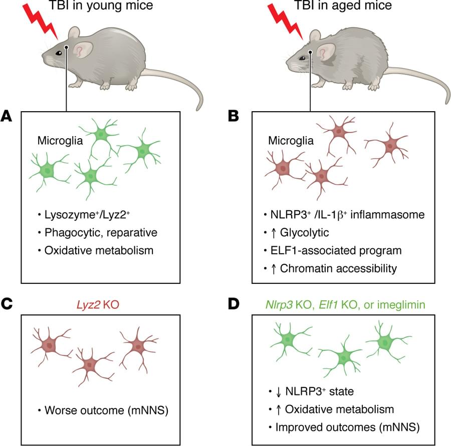
Bachstetter, Spinal Cord and Brain Injury Research Center, Department of Neuroscience, University of Kentucky, 741 S. Limestone Street, BBSRB Room B459, Lexington, Kentucky 40536–0509, USA. Phone: 859.218.4315; Email: [email protected].
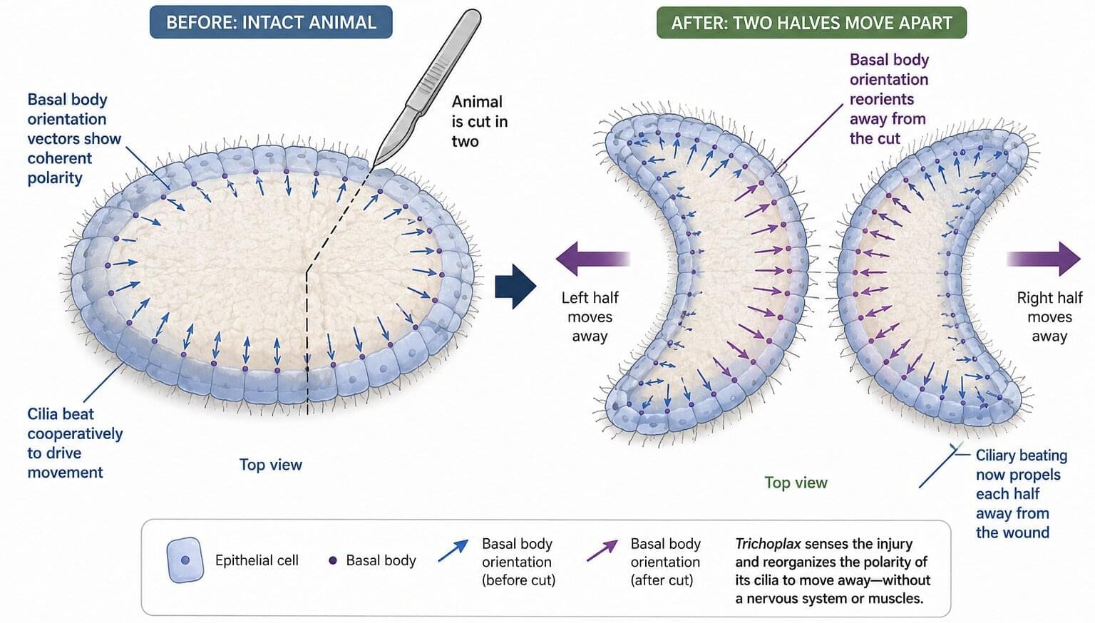
For a flat sea creature just a few millimeters across, a gentle poke is instantly recognized as danger. Trichoplax adhaerens—a translucent blob with no head, brain or muscles—scuttles away in seconds when touched. Imagine a flattened multicellular amoeba moving as a single unit: Trichoplax is only ~20 microns thick and a few millimeters wide. It glides on surfaces by beating tens of thousands of cilia on its lower epithelium (the underside), like microscopic oars dragging against the water.
Yet unlike most animals, Trichoplax has no obvious front or back end, no nerves or muscles at all. How can such a simple “crawling carpet” steer or change direction without a brain?
A new study reveals the remarkable flexibility of this pinhead-sized animal. While in most creatures, the orientation of each cilium is fixed early in development and locked to the body’s axes, Trichoplax achieves its swift escape by reorienting its thousands of hairlike cilia.