Casey Harrell uses his implants to talk to friends and family, read to his young daughter, and perform his job.
Category: neuroscience – Page 16
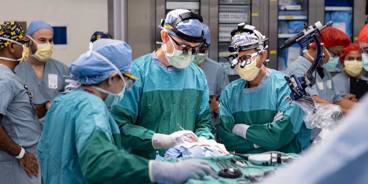
Paralyzed man walks again with help of ‘brain bridge’ implant
In a first of its kind procedure, a man left paralyzed after a spinal cord injury was able to walk again. Doctors implanted what they call a \.
What AI Reveals About the Brain
Can AI become smarter than humans?
In this episode, I talk to Chris Summerfield about the frontier of artificial intelligence, neuroscience, LLMs, AI agents, memory, and superintelligence.
We discuss why models like ChatGPT and Claude can feel so human, why today’s AI still does not learn like the brain, and why continual learning may be one of the most important unsolved problems in AI. Chris explains how human memory works, why sleep matters for learning, and what AI research is teaching us about intelligence itself.
We also discuss the future of work, education, creativity, and whether AI could lead to a more human world — or a much stranger one.
Topics covered:
• Artificial intelligence and the human brain.
• LLMs, ChatGPT, Claude and AI agents.
• AI memory and continual learning.
• AI alignment, safety and misalignment.
• . Superintelligence and self-improving systems.
• Hallucinations, reasoning and intelligence.
• . Education, jobs and the future of work.
• . Why AI may change how humans understand themselves.
TIMESTAMPS:
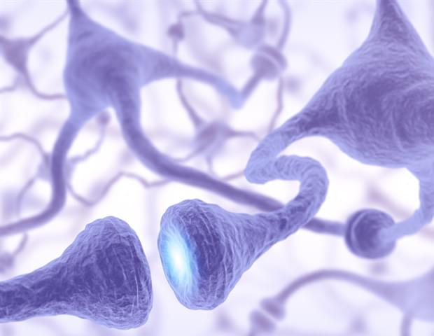
Scientists identify unique receptor that accelerates early neuron growth
Cells have surface receptors that couple to proteins and other molecules to initiate or inhibit certain behaviors. Typically, the number of these receptors increases as the cell matures, but researchers have now identified that one receptor influences cell behavior much earlier than previously thought and appears to help trigger the cell differentiation process to form neurons.
The Hiroshima University-based team published their work, which they said has implications for better understanding neuronal development and brain plasticity — and how those processes become dysregulated — on March 20 in iScience. They specifically found that G protein-coupled receptor 3 (GPR3) represents a unique molecule in this receptor family, as it behaves like an immediate-early gene that rapidly responds and induces downstream signaling. Other G protein-coupled receptors behave like delayed-response genes that aren’t expressed into much later in the cell maturation process.
Understanding early transcriptional responses — how genes are expressed in response to upstream signals — is critical because these programs determine neuronal development, synaptic formation and plasticity, and their dysregulation is associated with neurological disorders such as autism and cognitive dysfunction.
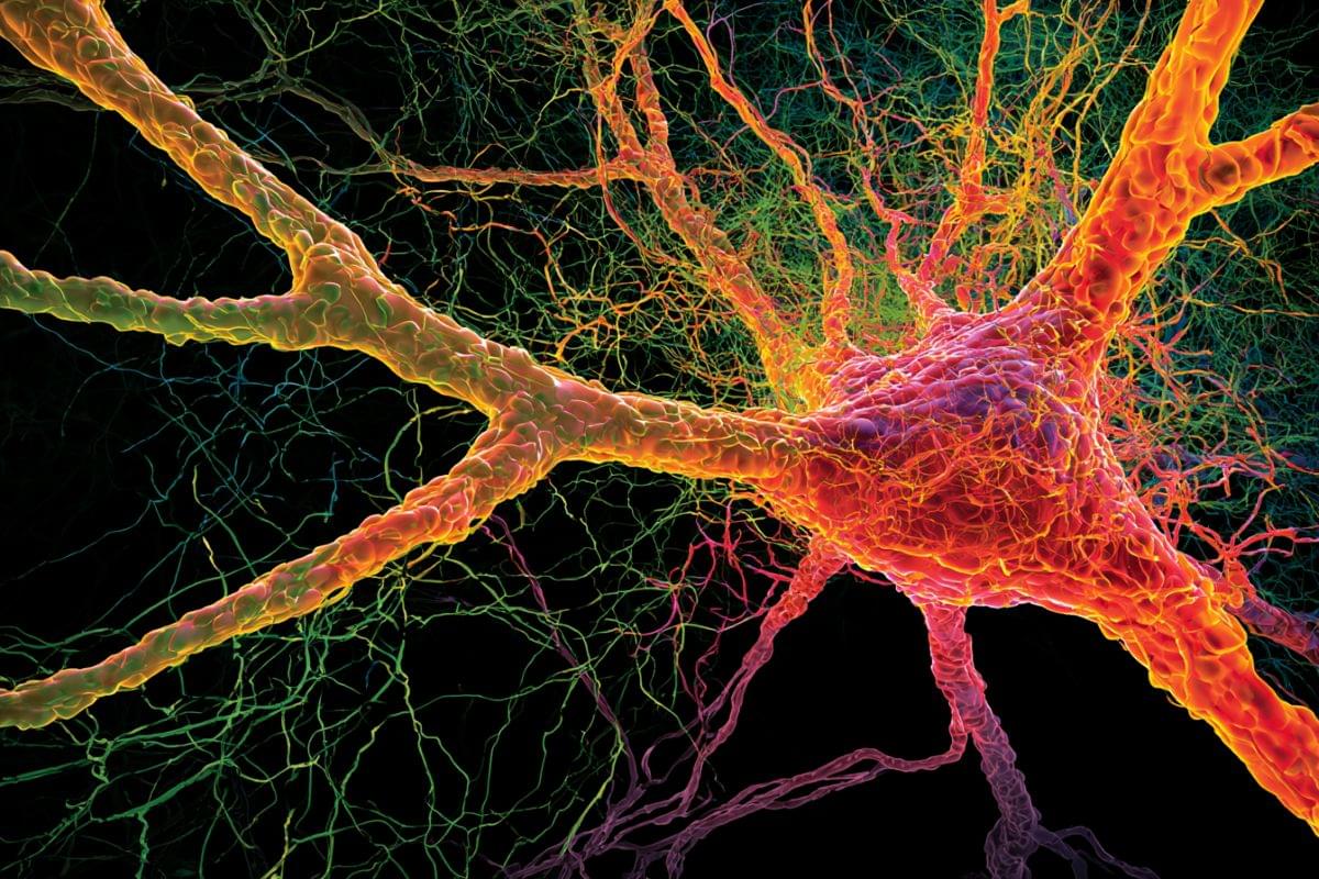
Alzheimer’s Protein APP Acts as Vital Shield for Neurons
Author: Hideaki Matsui Source: Niigata University Contact: Hideaki Matsui – Niigata University Image: The image is credited to Neuroscience News.
Original Research: Closed access. “A protective role for APP in nuclear waste clearance via lysosomal exocytosis” by Dougnon G, Otsuka T, Nakamura Y, Sakai A, Yamanaka T, Matsui N, Nakahara A, Ito A, Hatano A, Matsumoto M, Igarashi H, Kakita A, Ueno M, Matsui H. PNAS DOI:10.1073/pnas.
Abstract.
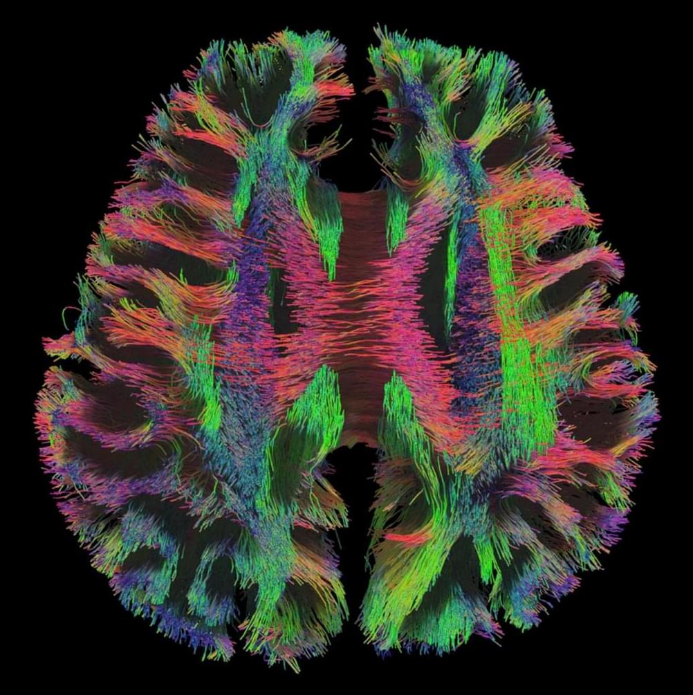
The Connectome 2.0 Scanner Captures the Brain in Unprecedented Detail
A one-of-a-kind MRI machine helps researchers see the relationship between the structure of the brain and how it functions.
Building Brains: The Molecular Logic of Neural Circuits
Thomas M. Jessel, Howard Hughes Medical Institute Investigator, explores the human brain, the sophisticated product of 500 million years of vertebrate evolution, assembled during just nine months of embryonic development. The functions encoded by its trillion nerve cells direct all human behavior. Yet the brain is a biological organ made from the same building blocks as skin, liver and lung. How does the brain acquire its remarkable computational power? Answers lie in the details of its construction — the cellular and molecular mechanisms that drive the formation of thousands of neural circuits, each wired for a specific behavior.
Developing brain cells routinely repair severe DNA damage during migration
Newborn nerve cells must squeeze through crowded, narrow spaces-through dense tissue, past other cells, between fibers-to reach the areas where they form neural circuits in the brain cortex.
In a new study published in Nature, researchers at Kyoto University’s Institute for Integrated Cell-Material Sciences (WPI-iCeMS) and their collaborators report that this journey causes widespread DNA damage in neurons, resulting in double-strand breaks where both strands of the double helix are completely severed. While this is the most severe type of DNA damage-capable of causing mutations and cell death-the team surprisingly found that it is a normal, routine feature of brain cortex formation, and a healthy brain quickly repairs it before harm occurs.
“The developing brain appears to have evolved to tolerate and repair the neuronal damage efficiently,” says Professor Mineko Kengaku, of WPI-iCeMS, who led the study. “But understanding the limits of that tolerance-and what happens when repair is incomplete-brings us closer to understanding a range of neurological conditions.”
Beyond Neuralink: How China’s Bio-Tech Breakthrough Fuels Next-Gen Brain-Computer Interfaces
From ultra-flexible materials redefining brain-computer interfaces (BCIs) to record-shattering global out-licensing deals, China’s biopharmaceutical sector is undergoing a profound qualitative transformation. ShanghaiEye takes you inside the Yunfan Future Factory and the cross-discipline innovation hub hosted by Chia Tai Tianqing (CTTQ)—a subsidiary of top-50 global pharma giant Sino Biopharmaceutical—to explore the cutting-edge ecosystem driving the future of global healthcare.
We examine a breakthrough BCI technology developed in Shanghai: an ultra-flexible photoresist material for neural electrode arrays. Ye Tianyang, CEO and Co-Founder of Yunfan Future, explains how this material—engineered to be 1,000 times softer than the rigid alternatives utilized by Western counterparts like Elon Musk’s Neuralink—exponentially reduces tissue damage and immune rejection. With dozens of human clinical trials already successfully completed worldwide, this innovation highlights the immense strength of Shanghai’s local talent pool and medical device supply chain.
The feature also spotlights the strategic roadmap of China’s pharmaceutical leaders. Eric Tse, CEO of Sino Biopharmaceutical and Chairman of CTTQ, breaks down their vision to build an open, interdisciplinary incubator. This global nexus bridges experts, scholars, and upstream and downstream partners, transforming Shanghai into a premier launchpad for international innovative drugs. Furthermore, Mr. Tse discusses the \.