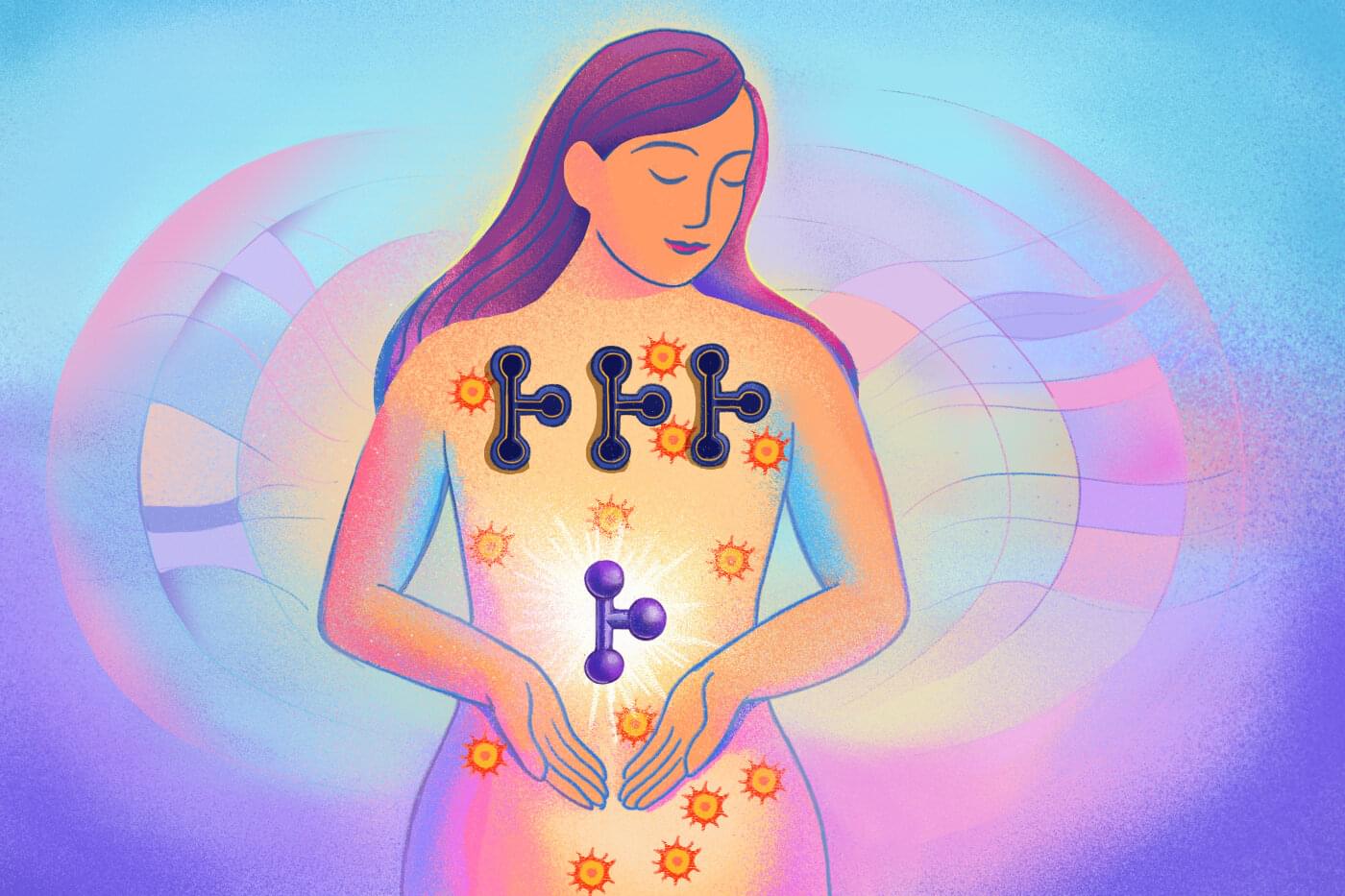
A hidden gut-brain protein may hold the key to stopping alcohol’s twin assault on the liver and the mind.

New in JNeurosci from Wei, Tao, Bi et al: Smokers who have quit their nicotine use have altered brain activity linked to heightened pain sensitivity and a need for more postoperative pain relief.
▶️
Perioperative abstinent smokers experience heightened pain sensitivity and increased postoperative analgesic requirements, likely due to nicotine withdrawal-induced hyperalgesia. However, the underlying neural mechanisms in humans remain unclear. To address this issue, this study enrolled 60 male patients (30 abstinent smokers and 30 nonsmokers) undergoing partial hepatectomy, collecting clinical data, smoking history, pain-related measures, and resting-state functional magnetic resonance imaging (rs-fMRI). Compared to nonsmokers, abstinent smokers showed lower pain threshold and higher postoperative analgesic requirements. Neuroimaging revealed altered brain function in abstinent smokers, including reduced fractional amplitude of low-frequency fluctuations (fALFF, 0.01 – 0.1 Hz) in the ventromedial prefrontal cortex (vmPFC), increased regional homogeneity (ReHo) in the left middle occipital gyrus, and decreased functional connectivity (FC) between the vmPFC to both the bilateral middle temporal gyrus and precuneus. Preoperative pain threshold was positively correlated with abstinence duration and specific regional brain activities and connectivity. Further, the observed association between abstinent time and pain threshold was mediated by the calcarine and posterior cingulate cortex activity. The dysfunction in vmPFC and left anterior cingulate cortex was totally mediated by the association between withdrawal symptoms and postoperative analgesic requirements. These findings suggest that nicotine withdrawal might alter brain functional activity and contribute to hyperalgesia for the abstinent smokers. This study provided novel insights into the supraspinal neurobiological mechanisms underlying nicotine withdrawal-induced hyperalgesia and potential therapeutic targets for postoperative pain in abstinent smokers.
Significance statement Abstinent smokers experienced heightened pain and require more analgesics after surgery, yet the underlying neural mechanisms remain poorly understood. This prospective cohort study identified altered regional brain activity associated with reduced pain thresholds and increased postoperative analgesic requirements in abstinent smokers. We found specific brain regions that were functionally altered and correlated with pain-related outcomes, which mediated the relationship between abstinence and pain-related behaviors. These findings provided novel insights into the supraspinal mechanisms of nicotine withdrawal-induced hyperalgesia and point to potential therapeutic targets for improving postoperative pain management in abstinent smokers.

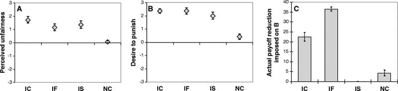
New brain research reveals why we’re willing to go out of our way to punish people who break the rules, even when it costs us time, money, or friends. This behavior, which researchers call “altruistic punishment,” has been essential for human cooperation since ancient times. It’s the invisible glue that keeps societies fair: we enforce the rules not just for ourselves, but for everyone.
Many people voluntarily incur costs to punish violations of social norms. Evolutionary models and empirical evidence indicate that such altruistic punishment has been a decisive force in the evolution of human cooperation. We used H2 15 O positron emission tomography to examine the neural basis for altruistic punishment of defectors in an economic exchange. Subjects could punish defection either symbolically or effectively. Symbolic punishment did not reduce the defector’s economic payoff, whereas effective punishment did reduce the payoff. We scanned the subjects’ brains while they learned about the defector’s abuse of trust and determined the punishment. Effective punishment, as compared with symbolic punishment, activated the dorsal striatum, which has been implicated in the processing of rewards that accrue as a result of goal-directed actions.

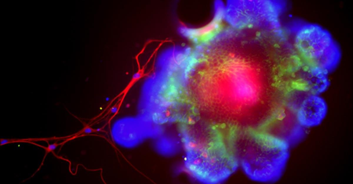
Cedars-Sinai researchers created “young” immune cells from human stem cells that reversed cognitive decline and Alzheimer’s symptoms in mice. The treated animals showed better memory and healthier brain structures. The cells seemed to protect the brain indirectly, possibly through anti-aging signals in the blood. The findings suggest a new, personalized path to slowing brain aging.
Northeastern University researchers have made a breakthrough drug discovery, developing the first synthetic endogenous cannabinoid compound, with repercussions for new therapeutics from pain and inflammation to cancer.
Spyros P. Nikas, an associate research professor in Northeastern’s Center for Drug Discovery, says that the discovery hinges on the distinction between two different kinds of cannabinoid chemicals, endogenous and exogenous. Exogenous cannabinoids are those produced outside the human body, like THC or CBD, both derived from the cannabis plant and present in marijuana.
Our own bodies, however, are also producing cannabinoids all the time. Called endogenous cannabinoids —or just “endocannabinoids”—these chemicals “modulate a wide range of physiological and pathophysiological responses,” Nikas says, processes that include mood, inflammation and even neurodegenerative disorders like Alzheimer’s and Parkinson’s. The research is published in the Journal of Medicinal Chemistry.

Question What are the main predictors for high health care costs among patients with head and neck cancer?
Findings In this population-based cohort study, advanced cancer stage and receiving multiple treatment modalities were the strongest predictors of high health care costs. Female sex, older age, and lower socioeconomic status were associated with an increased likelihood for high health care costs, although with a weaker effect size.
Meaning Future research should focus on evaluating screening strategies and early diagnosis to assess their potential effects on cost reduction and improved outcomes for patients with head and neck cancer.

A molecular switch has the ability to turn on a substance in animals that repairs neurological damage in disorders such as multiple sclerosis (MS), Mayo Clinic researchers discovered. The early research in animal models could advance an already approved Food and Drug Administration therapy and also could lead to new strategies for treating diseases of the central nervous system.
Research by Isobel Scarisbrick, Ph.D., published in the Journal of Neuroscience finds that by genetically switching off a receptor activated by blood proteins, named Protease Activated Receptor 1 (PAR1), the body switches on regeneration of myelin, a fatty substance that coats and protects nerves.
“Myelin regeneration holds tremendous potential to improve function. We showed when we block the PAR1 receptor, neurological healing is much better and happens more quickly. In many cases, the nervous system does have a good capacity for innate repair,” says Dr. Scarisbrick, principal investigator and senior author. “This sets the stage for development of new clinically relevant myelin regeneration strategies.”
Researchers have uncovered a fast, structural mechanism that allows neurons to stabilize communication when synaptic function is disrupted.
Instead of relying on electrical activity, the brain uses physical rearrangements of postsynaptic receptors to signal the sending neuron to boost neurotransmitter release.
This rapid correction restores balance within milliseconds, ensuring that circuits supporting movement, learning, and memory remain functional.
The findings shed new light on how the brain maintains stability when communication falters.
Neurons can rapidly rebalance their communication using a structural signal rather than electrical activity, overturning long-held assumptions about how synapses maintain stability.