Disclaimer: Do NOT attempt without proper medical supervision.
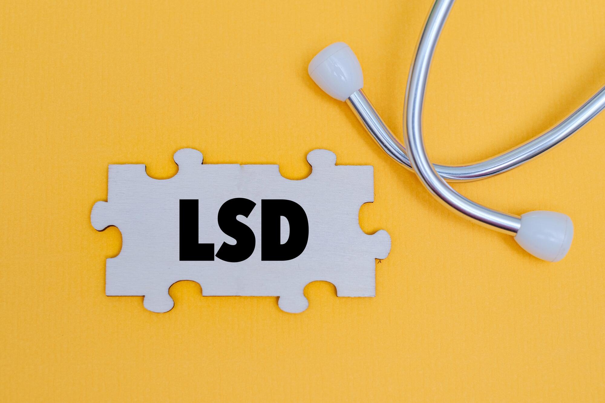
In a paradigm-shifting breakthrough, Phase III clinical trials of DT120 — a novel, pharmaceutical-grade formulation of LSD — have demonstrated unprecedented efficacy in treating Major Depressive Disorder (MDD) following just a single dose. The study, encompassing 149 patients, revealed that a one-time administration of DT120 significantly outperformed a placebo, achieving the trial’s primary endpoint by reducing MADRS depression scores by an 8-point margin at six weeks. Remarkably, patients experienced rapid therapeutic relief within just one week, showcasing a massive 14-point advantage over the placebo group. Unlike conventional daily antidepressants that often take weeks to manifest effects, DT120 delivers profound and sustained symptom reduction from a single intervention. Hailed by Definium Therapeutics’ CEO Rob Burrow as a potential “best-in-class” therapy, these groundbreaking findings not only pave the way for expedited regulatory approval but also underscore the transformative potential of psychedelics to fundamentally revolutionize modern mental health care.
Definium Therapeutics has announced strong results in a phase 3 trial of its single-dosed lysergide (LSD) drug DT120 in treating adults with major depressive disorder (MDD), meeting its primary goal and all key secondary efficacy endpoints in the first trial of its kind.
The results come from the Emerge trial, a randomized, double-blind, placebo-controlled study featuring 149 participants aged 18 to 74 years enrolled across 20 sites. The participants all met specific MDD measures. They needed to have a DSM-5-confirmed diagnosis of MDD, a Montgomery-Åsberg Depression Rating Scale (MADRS) score of at least 26 and a Clinical Global Impression–Severity (CGI-S) score of at least 4 at screening and baseline.
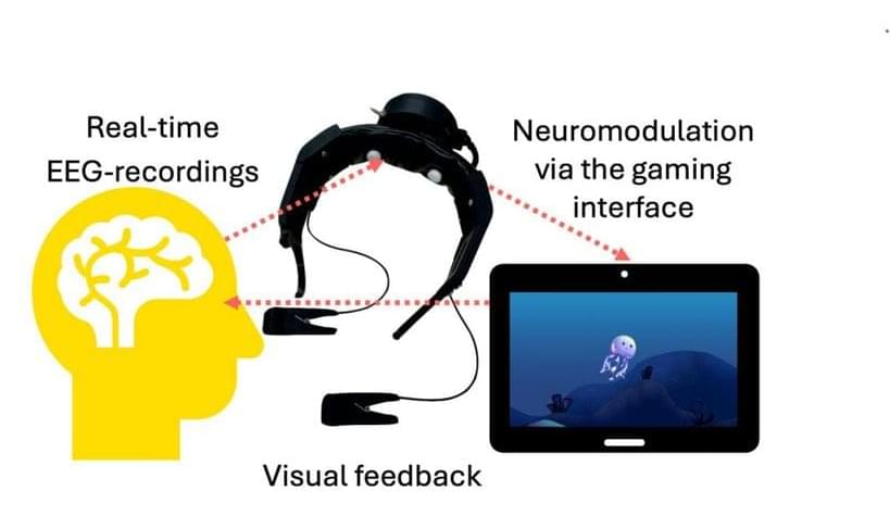
The study examined the effectiveness of a single 100 µg dose of DT120 ODT compared with a placebo in alleviating MDD symptoms. In 2023, we covered an earlier trial of lysergide, which had shown positive results in treating general anxiety disorder (GAD).

