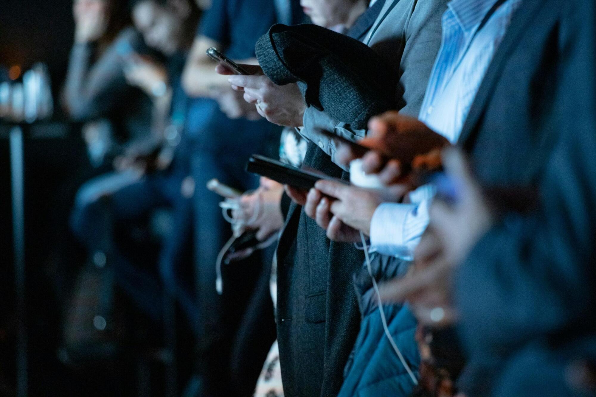
Toggle light / dark theme

Frontiers: Information storage and transfer in the brain require a high computational power

Neuronal network display various local or global mechanisms to allow information storage and transfer in the brain. From synaptic to intrinsic plasticity, the rules of input–output function modulation have been well characterized in neurons. In the past years, astrocytes have been suggested to increase the computational power of the brain and we are only just starting to uncover their role in information processing. Astrocytes maintain a close bidirectional communication with neurons to modify neuronal network excitability, transmission, axonal conduction, and plasticity through various mechanisms including the release of gliotransmitters or local ion homeostasis. Astrocytes have been significantly studied in the context of long-term or short-term synaptic plasticity, but this is not the only mechanism involved in memory formation. Plasticity of intrinsic neuronal excitability also participates in memory storage through regulation of voltage-gated ion channels or axonal morphological changes. Yet, the contribution of astrocytes to these other forms of non-synaptic plasticity remains to be investigated. In this review, we summarized the recent advances on the role of astrocytes in different forms of plasticity and discuss new directions and ideas to be explored regarding astrocytes-neuronal communication and regulation of plasticity.

The rules governing changes in synaptic and intrinsic plasticity are diverse and complex, sometimes synergistic and sometimes not (Debanne et al., 2019). Most studies have been neuro-centric, despite growing evidence that astrocytes can intervene or interact to modify or modulate synaptic transmission (Araque et al., 1998; Jourdain et al., 2007; Bonansco et al., 2011), input integration, neuronal excitability (Tan et al., 2017), spike waveform or axonal conductivity (Sasaki et al., 2011; Lezmy et al., 2021). Astrocytes can detect neuronal activity, and depending on the firing rate of action potentials (APs), they can not only release gliotransmitters such as adenosine or glutamate (Hamilton et al., 2008; Lezmy et al., 2021), but also trigger intracellular calcium ([Ca2+]i) oscillations at different frequencies (Pasti et al., 1997).

Protein modification in neurodegenerative diseases

The graphical abstract showcases the diversity of posttranslational modifications (PTMs) influencing protein structure and function. It features schematic representations of the following 10 prominent PTMs: phosphorylation (addition of phosphate groups), acetylation (addition of acetyl groups), methylation (addition of methyl groups), SUMOylation (attachment of SUMO proteins), ubiquitylation (attachment of ubiquitin molecules), succinylation (addition of succinyl groups), S-nitrosylation (attachment of NO), ADP-ribosylation (addition of ADP-ribose groups), glycosylation (addition of sugar molecules), and palmitoylation (attachment of palmitate groups).

Locus coeruleus–amygdala circuit disrupts prefrontal control to impair fear extinction

One of the most-viewed PNAS articles in the last week is “Locus coeruleus–amygdala circuit disrupts prefrontal control to impair fear extinction.” Explore the article here: https://ow.ly/yFH250Ywubb.

For more trending articles, visit https://ow.ly/tZsG50Ywubg.


Stress undermines extinction learning and hinders exposure-based clinical therapies for a variety of neuropsychiatric disorders. In both animals and humans, dysfunction in the ventromedial prefrontal cortex (vmPFC) contributes to stress-impaired extinction, but the neural circuit by which stress modulates vmPFC function is not known. We hypothesize that locus coeruleus (LC) norepinephrine undermines extinction learning by recruiting projections from the basolateral amygdala (BLA) to vmPFC. Using a combination of circuit-specific chemogenetics and calcium imaging, we find that activation of LC noradrenergic neurons mimics a behavioral stressor (footshock), induces freezing behavior, reduces spontaneous neuronal activity in the vmPFC, impairs extinction learning, and alters the population dynamics of vmPFC ensembles.

Survey: What are neuroscience’s most transformative new tools?

A nicely organized list of what various investigators highlight as the most transformative neuroscience tools from the past 5 years!


Which new tools—including artificial intelligence, deep-learning methods, genetic tools and advanced neuroimaging—are making the largest impact?

Using mRNA to Fight Tau Aggregation in Alzheimer’s

Researchers publishing in Cell Reports Medicine have described the development of a lipid nanoparticle (LNP) that delivers mRNA to neurons in order to stop the formation of tau aggregates and fight Alzheimer’s disease.

Tau and amyloids

Amyloid beta deposition between neurons and tau aggregation within neurons are both hallmarks of Alzheimer’s disease, and evidence suggests that the latter is potentially more significant than the former [1]. While some potential therapies have been discovered that may disaggregate these tau tangles after they have formed [2], no therapy has yet been approved by the FDA to do this.

The Janus face of NK cells in neurodevelopment

NK cells in neurodevelopment.

Maternal immune activation (MIA) during pregnancy perturbs fetal neurodevelopment, with natural killer (NK) cells emerging as key contributors to neurodevelopmental disorders (NDDs) such as autism spectrum disorder (ASD).

Clinical studies consistently report NK cell dysfunction in ASD patients and their mothers, characterized by altered cytotoxicity, hyperactivation at rest, functional exhaustion on stimulation, and skewed receptor/genetic profiles.

Uterine NK (uNK) cells, indispensable for placental and fetal development, can paradoxically promote NDDs when hyperactivated, releasing granzyme B (GZMB) that disrupts fetal brain structure and function.

Elucidating the MIA-driven ‘uNK/ GZMB–microglia–NDD’ axis is essential to devise preventive strategies for high-risk pregnancies and identify early biomarkers of neurodevelopmental risk. sciencenewshighlights ScienceMission https://www.cell.com/cms/10.1016/j.it.2025.10.001/asset/89cd…ts/gr3.jpg https://sciencemission.com/Janus-face-of-NK-cells


Maternal immune activation (MIA), triggered by infection or inflammation during pregnancy, is a well-recognized risk factor for neurodevelopmental disorders (NDDs) such as autism spectrum disorder (ASD). Clinical cohort studies and rodent models suggest that natural killer (NK) cells play a significant role in NDD pathogenesis, but the underlying mechanisms remain poorly defined. Here, we summarize the key immune mediators involved in MIA-induced NDDs, emphasizing microglia as a central hub. We then examine emerging evidence implicating aberrant NK cell activation in ASD, underscoring their overlooked contribution to impaired neurodevelopment. Finally, we discuss potential mechanisms of NK cell–microglia crosstalk in NDDs. Elucidating these interactions in the context of MIA will be crucial for developing preventive and therapeutic strategies against inflammation-driven NDDs.

Biological and Clinical Staging of Alzheimer Disease Pathology in Down Syndrome

Among adults with DownSyndrome, clinical and biological staging of AlzheimerDisease showed greater concordance compared to sporadic cases, supporting the use of cognition-based staging for clinical trial enrollment. Most discordant cases exhibited more advanced pathology than clinical stage, highlighting resilience mechanisms in this population.


This cross-sectional study examines data for participants in the Alzheimer Biomarker Consortium–Down Syndrome study to gauge the concordance of clinical and biological Alzheimer disease staging.

A direct auditory subcortical route to the amygdala associated with fear in humans

New in JNeurosci from Kosteletou-Kassotaki et al: A white matter tract connecting the inferior colliculus to the basolateral amygdala via the MGB of the thalamus is linked to better hearing ability and higher self-reported fearfulness in people.

▶️


Rapid and efficient fear processing is essential for survival. In vision, this function is supported by a well-characterized subcortical pathway consisting of direct projections from the pulvinar of the thalamus to the amygdala in the human brain. In contrast, the existence of an analogous shortcut for fear in audition has been demonstrated in non-human animals, but remains unconfirmed in humans. To address this question, we used probabilistic streamline tractography and fixel-based analysis on diffusion-weighted images from Human Connectome Project participants of either sex, to reconstruct candidate auditory subcortical pathways and examine their associations with affective and auditory behavioral measures. Our findings revealed a robust white matter tract connecting the inferior colliculus to basolateral amygdala via the medial geniculate body (MGB) of the thalamus. Remarkably, higher fiber density in this tract was associated with better hearing ability in noise and increased self-reported fearfulness, supporting its role in auditory and affective function. Conversely, a control analysis of the core thalamocortical pathway from ventral MGB to primary auditory cortex (PAC), representing the main route for auditory processing, was associated with auditory ability but not with affective measures. These findings provide previously unreported evidence for an auditory colliculo-geniculo-amygdala “low road” in humans, aligning with evolutionarily conserved pathways for fear described in non-human species.

Significance Statement Rapid fear processing is crucial for survival. While a visual subcortical “low road” for fear is well characterized in humans, the existence of an analogous human auditory shortcut remains undetermined. Using diffusion magnetic resonance imaging tractography, we provide evidence for a white matter tract connecting the inferior colliculus to basolateral amygdala via the medial geniculate body of the thalamus, which is associated with hearing ability and self-reported fearfulness. Our findings provide novel evidence for an auditory colliculo-geniculo-amygdala direct route in humans, revealing an evolutionarily conserved pathway for fear previously described in non-human species.

GLP-1 medications used to treat diabetes and obesity may also help alleviate symptoms of anxiety and depression

GLP-1 medications used to treat diabetes and obesity were associated with a reduced need for hospital care and sickness absence due to psychiatric reasons, a new study shows. The large register-based study was carried out in collaboration between the University of Eastern Finland, Karolinska Institutet in Stockholm and Griffith University in Australia.

Diabetes and obesity are associated with an increased risk of mental health symptoms, and similarly, individuals with mental disorders have an elevated risk of metabolic diseases such as obesity and diabetes. Researchers have long been interested in the connections between these conditions and in how pharmacological treatments may affect both metabolic and mental health disorders.

The present study included nearly 100,000 participants, more than 20,000 of whom had used GLP-1 medications. Participants were followed through Swedish national registers between 2009 and 2022. The study’s findings were published in The Lancet Psychiatry.

/* */