Among participants with EUR-like genomes, SNP heritability (h2SNP) estimated for the Big Five traits using linkage disequilibrium score regression (LDSC)34 ranged from 4.8% (s.e. = 0.2%) for agreeableness to 9.3% (s.e. = 0.3%) for extraversion (Table 1, Supplementary Table 4 and Supplementary Note 3). Importantly, these SNP heritability estimates from GWAS meta-analysis index genetic effects that are consistent across contributing cohorts. To allow for variability in genetic effects across cohorts, we conducted a random effects meta-analysis of cohort-specific h2SNP estimates, which indicated an average h2SNP of 8.6% (s.e. = 0.6%) across traits (ranging from 7.4% for agreeableness to 10.6% for extraversion; Table 1 and Supplementary Table 21), with significant variability across cohorts (mean τ = 3.6%). Random response error by the participants cannot systematically relate to their genome35,36. Accordingly, we found that personality measures with greater reliability (lower random response error) tended to be more heritable (b = 6.7%, s.e. = 0.7%; Extended Data Fig. 2). In this analysis, the expected h2SNP for a measure of typical (median) reliability (α = 0.81) ranged from 9.3% for agreeableness (s.e. = 0.6%) to 13.3% for extraversion (s.e. = 0.6%), and h2SNP completely disattenuated for measurement error ranged from 10.8% for agreeableness (s.e. = 0.9%) to 15.8% for extraversion (s.e. = 0.9%; Table 1).
To further characterize the generalizability of genetic associations with personality, we examined the concordance of genetic signal across geography, age, veteran status, measurement instrument and reporter perspective (Table 1 and Extended Data Fig. 3). Genetic effects were similar but not identical across four western country clusters (USA, continental Europe, Nordic and UK–Australia, mean rg = 0.86, mean s.e. = 0.15), three age groups (young (≤25 years), middle (25–64 years) and older (65 years and older), mean rg = 0.80, mean s.e. = 0.18), between the Million Veteran Program and other, primarily non-veteran cohorts (mean rg = 0.82, mean s.e. = 0.04), and across five personality measurement instruments (mean rg = 0.85, mean s.e. = 0.07). Additional characterization of genetic architecture across measurement instruments using genomic structural equation modelling37 confirmed that genetic effects plausibly operate at the level of broad cross-instrument latent factors, with only one locus showing significantly heterogenous effects across measurement instruments (Supplementary Tables 22 – 24 and Extended Data Fig. 4). Notably, genetic associations with agreeableness were less consistent across cohorts (Table 1), explaining in part why agreeableness exhibited lower heritability than other traits in the meta-analytic GWAS. In the Estonian Biobank, in which the personality of the participants was assessed both by their self-report (n = 73,983) and by reports by close others (n = 20,269), we found strong genetic overlap between rater perspectives (mean rg = 0.84, mean s.e. = 0.12), indicating that the genetic architecture of personality is not an epiphenomenon of self-perception. In sex-stratified analyses of neuroticism in the UK Biobank cohort, X-chromosome-linked h2SNP did not differ between male individuals (n = 168,989; h2SNP, X = 0.23%; s.e. = 0.04%) and female individuals (n = 198,139; h2SNP, X = 0.18%; s.e. = 0.03%; Pdifference = 0.33). The dosage compensation ratio (\(\hat{{m{\gamma }}}\) = 1.26, s.e. = 0.30) was intermediate between no compensation (0.5) and full compensation (2.0) but was estimated relatively imprecisely. Genetic effects were correlated near-unity across sex (rg = 0.96; 95% confidence interval (CI) = 0.81–1.10).
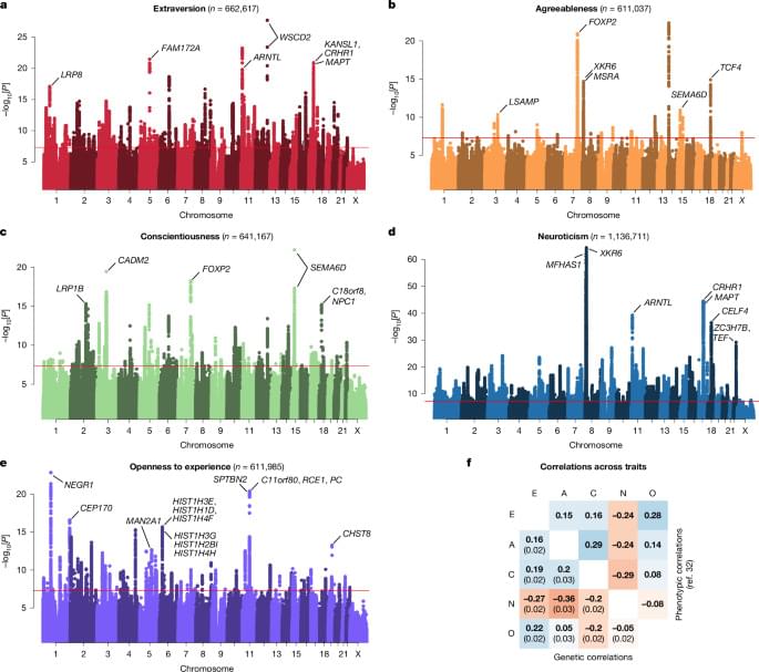
Biological follow-up of GWAS signals indicated that enriched gene sets intersected across the Big Five (mean enrichment rank-order ρ = 0.72; Extended Data Fig. 5), providing evidence for trait-overlapping molecular and cellular systems in personality neurobiology despite only modest genetic correlations (Fig. 1f). Consistent with theories of personality development that emphasize the prefrontal cortex38,39, genetic associations for each Big Five trait, except for agreeableness, were enriched in genes expressed in the prefrontal cortex (among these, top lead SNPs implicate RCE1, FOXP2 and SEMA6D, indicated in Fig. 1; Supplementary Tables 25–34). All traits demonstrated strong enrichment in protein-truncating variant-intolerant gene sets specifically expressed in neurons (such as ARNTL, TCF4 and NEGR1; Fig. 1).