Learn more about how younger generations are now aging faster than their parents and grandparents, and how this aging gap could explain the rise in early-onset cancers.

Can aging be reversed? Dr. Michael West explains telomerase, cellular immortality, stem cells, tissue regeneration, and the future of longevity.
LifeCraft Sciences:
https://lifecraftsciences.com/
In this episode, I sit down with pioneering molecular gerontologist and biotechnology entrepreneur Dr. Michael D. West to explore telomeres, telomerase, cellular senescence, stem cells, tissue regeneration, and the possibility of reversing biological aging.
One of our central topics is the groundbreaking telomerase program West founded and led at Geron. That research helped establish how restoring telomerase activity can protect the ends of chromosomes and allow normal human cells to move beyond their usual replicative limit while retaining youthful characteristics in laboratory culture. We unpack what scientists mean when they say a cell has been “immortalized,” why cellular immortality is very different from making a person immortal, and how telomerase connects the biology of aging with the biology of cancer.
We also explore West’s work in regenerative medicine and his early vision of pluripotent stem cells as a “parts supply store” for the human body. Could youthful cells eventually be used to repair damaged tissues, replace worn-out biological components, and restore regenerative capabilities lost with age? West discusses the early isolation of human embryonic stem cells, therapeutic cloning, developmental reprogramming, and what cloned animals taught researchers about resetting cellular age.
Finally, we discuss LifeCraft Sciences and RESTORE, the company’s experimental approach combining telomerase with developmental regulators to return aged cells to a more youthful, regenerative state. It is a fascinating conversation about the history of longevity science, the future of tissue repair, and one of biology’s biggest questions: can aging eventually be reversed rather than merely slowed?
What if aging isn’t just biology…but also psychology — and your brain is quietly shaping how fast you age every day?
Dr. Srini Pillay, MD (https://drsrinipillay.com/) is a Harvard-trained psychiatrist, brain researcher, entrepreneur, author, and expert in the science of human potential, resilience, and longevity.
Dr. Pillay previously served as Assistant Professor of Psychiatry at Harvard Medical School and directed both the Outpatient Anxiety Disorders Program and the Panic Disorders Research Program in Brain Imaging at McLean Hospital, one of the world’s leading psychiatric institutions.
Over the course of his career, Dr. Pillay has focused on understanding how the brain shapes performance, creativity, emotional health, leadership, and even biological aging. His work bridges neuroscience, psychiatry, technology, and human behavior — translating cutting-edge brain science into practical tools for individuals, organizations, and healthcare systems.
Dr. Pillay is the co-founder and Chief Medical Officer of Reulay (https://www.reulay.com/), an AI-driven digital therapeutics and mindset technology company focused on healthy longevity, stress reduction, and human performance. He is also founder of the NeuroBusiness Group (https://nbgcorporate.com/), where he works with leaders and organizations around the world on brain-based approaches to innovation, adaptability, resilience, and navigating complexity in the age of AI.
Dr. Pillay is the author of several influential books including Tinker Dabble Doodle Try (https://www.amazon.com/Tinker-Dabble-?tag=lifeboatfound-20… which explores the neuroscience of creativity and the untapped power of the brain’s unconscious processing systems.
She says she reversed 20 years of biological aging by testing gene therapy on her own body before anyone else. In this episode of The 200 Year Life Project, Gary Leland sits down with Elizabeth Parrish, founder and CEO of BioViva, the first person to undergo gene therapy specifically aimed at reversing aging.
Gary, 71 and dead serious about reaching 200, talks with Parrish about how telomere and myostatin gene therapy works, what her published telomere data showed, why these therapies are still done outside the US, and how she believes affordable gene therapy could change human lifespan and healthspan. They also get into longevity escape velocity, the \.
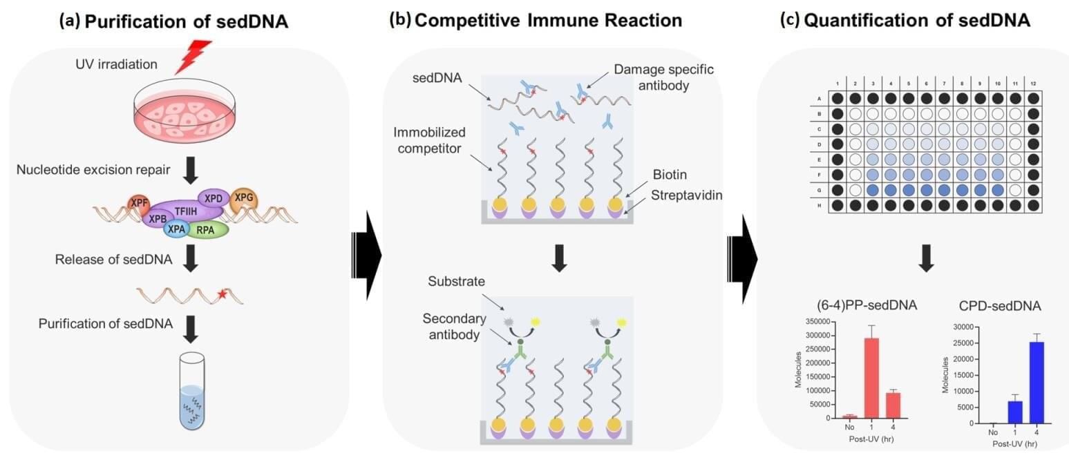
The Korea Research Institute of Standards and Science has developed an ultrasensitive immunoassay-based analytical platform that can detect and quantify trace amounts of “Small Excised Damaged DNA (sedDNA)” fragments generated during cellular DNA repair. This technology enables highly sensitive detection with quantification down to the level of several thousand molecules, measuring up to 22 times more DNA fragments than conventional methods. It provides a new analytical foundation for comparing DNA repair capacity between individuals and studying cellular responses to anticancer drugs and carcinogenic agents.
Human DNA is continuously exposed to damage from ultraviolet light, chemical agents, smoking and normal metabolic processes. If such damage is not properly repaired, mutations can accumulate and lead to aging and diseases such as cancer. To maintain genomic stability, cells activate the Nucleotide Excision Repair (NER) system, which removes damaged DNA segments and replaces them with newly synthesized DNA. The small excised DNA fragments generated during this process serve as important indicators of DNA repair efficiency and kinetics, providing a valuable tool for studying disease mechanisms and predicting treatment responses.
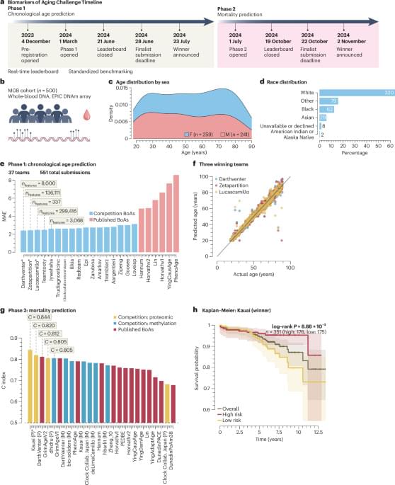
Ying, K., Paulson, S., Reinhard, J. et al. An open competition for biomarkers of aging. Nat Aging 6, 1193–1195 (2026). https://doi.org/10.1038/s43587-026-01139-6
The retina of the human eye contains 6–7 million cone cells. These cells contain light-sensitive proteins known as cone opsins. They enable us to perceive our surroundings in detail in daylight. They allow us to see the world in thousands of colors: red strawberries, green leaves, the blue sky. They also enable us to see all the objects around us clearly. And they allow us to perceive fast movements, such as the rush of a train or the flight of a dragonfly.
Often, however, these all-rounders of daylight vision are also involved in retinal diseases. Impairment of cone receptor function, caused by genetic mutations or other degenerative processes, can lead to disorders such as color blindness and age-related macular degeneration (AMD), a disease affecting the central retina and causing progressive vision loss.
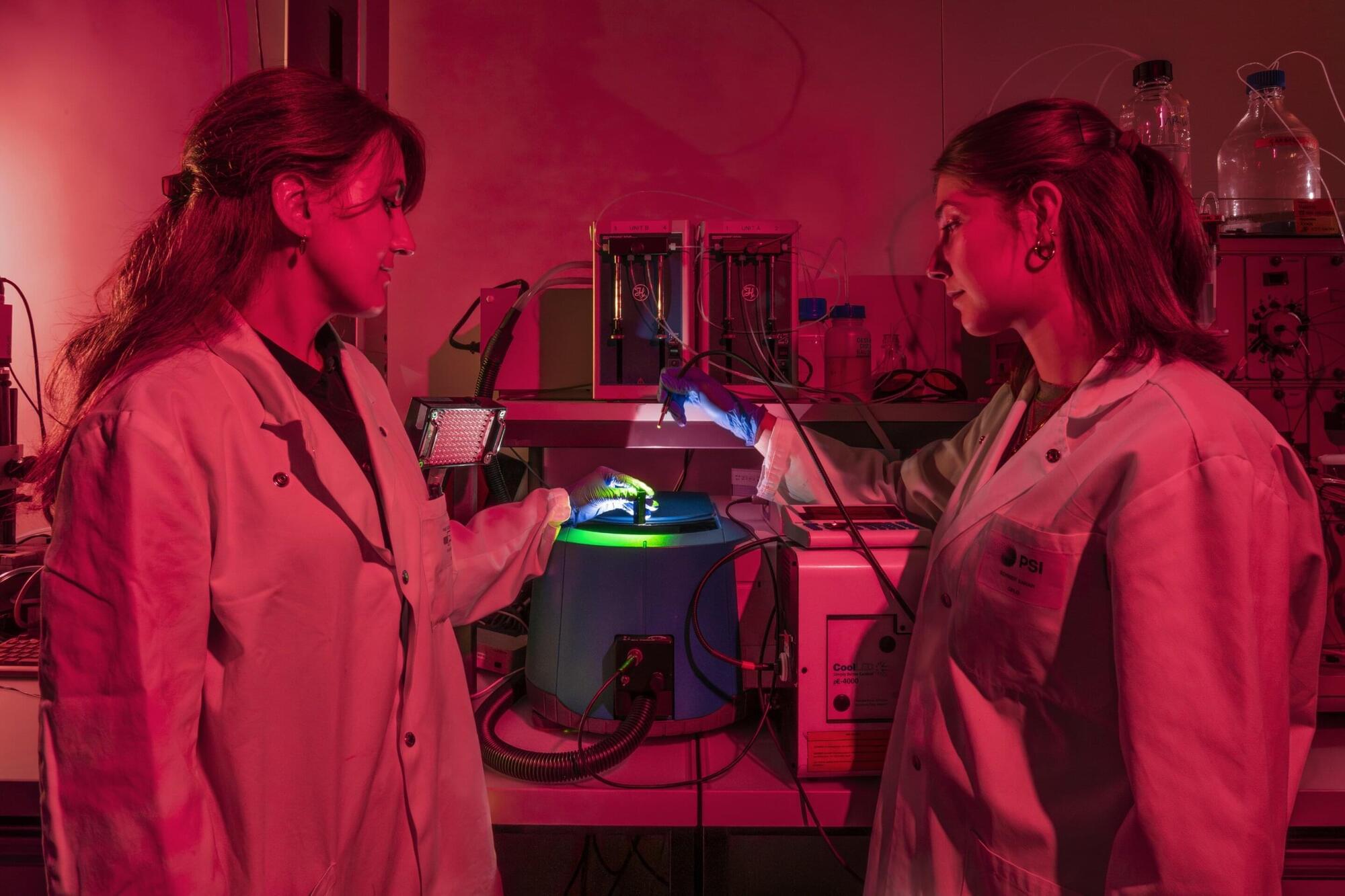
In a new study, Polina Isaikina and Sarah L. Schmidt, two researchers from the Center for Life Sciences at PSI, have succeeded for the first time in determining the three-dimensional structure of human cone opsins in their dark state and showing how their molecular architecture enables their rapid activation by light.
Awesome results and a new project to double mice lifespan. If I could fund one researcher right now it would be this man.
In this Conference talk, Dr. Greg Fahy presents stunning data from the TRIIM and TRIIM-X trials. His team has successfully regrown the human thymus in older adults, reversed epigenetic aging clocks by up to two years, and restored immune function to levels seen decades earlier.
Beyond the lab results, participants showed dramatic real-world improvements: 15% stronger muscles, 21% better VO2 max, and frailty scores dropping to near zero. Dr. Fahy also unveils the \.