Toggle light / dark theme

The breakthrough marks a promising target for drug therapies that slow, possibly reverse, the disease’s development

NEW YORK, NY, December 23, 2024 — Researchers with the CUNY ASRC have unveiled a critical mechanism that links cellular stress in the brain to the progression of Alzheimer’s disease (AD). The study, published in the journal Neuron, highlights microglia, the brain’s primary immune cells, as central players in both the protective and harmful responses associated with the disease.

Microglia, often dubbed the brain’s first responders, are now recognized as a significant causal cell type in Alzheimer’s pathology. However, these cells play a double-edged role: some protect brain health, while others worsen neurodegeneration. Understanding the functional differences between these microglial populations has been a research focus for Pinar Ayata, the study’s principal investigator and a professor with the CUNY ASRC Neuroscience Initiative and the CUNY Graduate Center’s Biology and Biochemistry programs.

ZMQ-1, a novel aluminosilicate zeolite with interconnected meso-microporous channels, addresses limitations of traditional zeolites by enhancing stability and catalytic efficiency.

Researchers have developed a groundbreaking aluminosilicate zeolite, ZMQ-1, designed with a distinctive intersecting meso-microporous channel system. This innovation is poised to significantly improve catalytic processes in the petrochemical industry.

Published in Nature, the study presents ZMQ-1 as the first aluminosilicate zeolite featuring interconnected intrinsic 28-ring mesopores. This breakthrough addresses long-standing challenges in zeolite design, including limitations in pore size, stability, and catalytic efficiency.

Researchers at Australia’s Monash University are using a common medicine cabinet antiseptic in unique battery chemistry that could soon power drones and other electric aircraft, according to a school news release.

The team is tapping Betadine, a common brand name for a topical medication used to treat cuts and other wounds, in research garnering surprising results.

“… We found a way to accelerate the charge and discharge rates, making them a viable battery option for real-world heavy-duty use,” paper first author and doctoral student Maleesha Nishshanke said in the release.

Understanding the behavior of the molecules and cells that make up our bodies is critical for the advancement of medicine. This has led to a continual push for clear images of what is happening beyond what the eye can see. In a study recently published in Science Advances, researchers from Osaka University have reported a method that gives high-resolution Raman microscopy images.

Raman microscopy is a useful technique for imaging because it can provide about specific molecules—such as proteins—that take part in the body’s processes. However, the Raman light that comes from biological samples is very weak, so the signal can often get swamped by the background noise, leading to poor images.

The researchers have developed a microscope that can maintain the temperature of previously frozen samples during the acquisition. This has allowed them to produce images that are up to eight times brighter than those previously achieved with Raman microscopy.

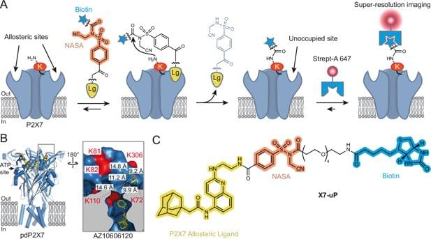
Quantum sensing is a rapidly developing field that utilizes the quantum states of particles, such as superposition, entanglement, and spin states, to detect changes in physical, chemical, or biological systems. A promising type of quantum nanosensor is nanodiamonds (NDs) equipped with nitrogen-vacancy (NV) centers. These centers are created by replacing a carbon atom with nitrogen near a lattice vacancy in a diamond structure.

When excited by light, the NV centers emit photons that maintain stable spin information and are sensitive to external influences like magnetic fields, electric fields, and temperature. Changes in these spin states can be detected using optically detected (ODMR), which measures fluorescence changes under .

In a recent breakthrough, scientists from Okayama University in Japan developed nanodiamond sensors bright enough for bioimaging, with spin properties comparable to those of bulk diamonds. The study, published in ACS Nano, on 16 December 2024, was led by Research Professor Masazumi Fujiwara from Okayama University, in collaboration with Sumitomo Electric Company and the National Institutes for Quantum Science and Technology.

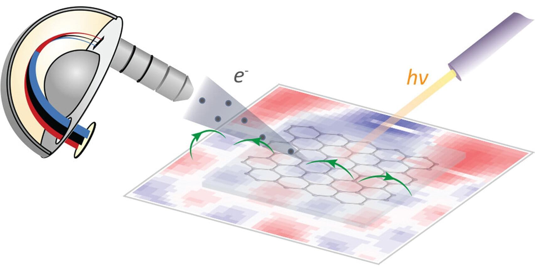
At the Berlin synchrotron radiation source BESSY II, the largest magnetic anisotropy of a single molecule ever measured experimentally has been determined. The larger a molecule’s anisotropy is, the better suited it is as a molecular nanomagnet. Such nanomagnets have a wide range of potential applications, for example, in energy-efficient data storage.

Researchers from the Max Planck Institute for Kohlenforschung (MPI KOFO), the Joint Lab EPR4Energy of the Max Planck Institute for Chemical Energy Conversion (MPI CEC) and the Helmholtz-Zentrum Berlin were involved in the study.

The research involved a bismuth complex synthesized in the group of Josep Cornella (MPI KOFO). This molecule has unique magnetic properties that a team led by Frank Neese (MPI KOFO) recently predicted in . So far, however, all attempts to measure the magnetic properties of the bismuth complex and thus experimentally confirm the theoretical predictions have failed.

The melting point is one of the most important measurements of material properties, which informs potential applications of materials in various fields. Experimental measurement of the melting point is complex and expensive, but computational methods could help achieve an equally accurate result more quickly and easily.

A research group from Skoltech conducted a study to calculate the maximum of a high-entropy carbonitrides—a compound of titanium, zirconium, tantalum, hafnium, and niobium with carbon and nitrogen.

The results published in the Scientific Reports journal indicate that high-entropy carbonitrides can be used as promising materials for protective coatings of equipment operating under —high temperature, thermal shock, and chemical corrosion.

Researchers discovered that the mRNA modification m6A triggers rapid degradation, regulating protein production. This breakthrough could inform drug development to manage protein-related diseases.

Messenger ribonucleic acids (mRNA) are like the architects of our bodies. They carry precise blueprints for building proteins, which are read and assembled by their cellular partners, the ribosomes. Proteins are essential for our survival, as they regulate cell division, bolster the immune system, and make our cells resilient against external threats.

Just like in real-world construction, some cellular blueprints require extra instructions—such as when a protein needs to be produced rapidly or when corrections are needed for a flawed design. In our bodies, this role is fulfilled by RNA modifications. These small chemical changes function like detailed annotations, offering additional guidance to specific parts of the mRNA for optimal protein production.

A recent study from Stanford’s Wu Tsai Neurosciences Institute has shed light on the interplay between two key brain chemicals, dopamine and serotonin, revealing their opposing roles in shaping our decisions and learning processes. Published in Nature, the research demonstrates for the first time that dopamine and serotonin operate as a “gas and brake” system, jointly influencing how we learn from rewards. The findings have broad implications, from understanding everyday decision-making to developing treatments for neurological and psychiatric conditions such as addiction, depression, and Parkinson’s disease.

Dopamine and serotonin are crucial to many aspects of human behavior, including reward processing and decision-making. Both neurotransmitters are also implicated in a variety of mental health disorders. While previous research has established their individual roles—dopamine is linked to reward prediction and seeking, while serotonin promotes long-term thinking and patience—the precise nature of their interaction has remained unclear.

Two competing theories have sought to explain their dynamic: the “synergy hypothesis,” which posits that dopamine focuses on immediate rewards and serotonin on long-term benefits, and the “opponency hypothesis,” suggesting the two act in opposition, with dopamine encouraging impulsive action and serotonin promoting restraint. The Stanford researchers aimed to directly test these theories using advanced experimental methods.

Researchers from Tokyo Metropolitan University have made tungsten disulfide nanotubes which point in the same direction when formed, for the first time. They used a sapphire surface under carefully controlled conditions to form arrayed tungsten disulfide nanotubes, each consisting of rolled nanosheets, using chemical vapor deposition.

The team’s technique resolves the long-standing issue of jumbled orientations in collected amounts of nanotubes, promising real world applications for the exotic anisotropy of single nanotubes.

The study is published in the journal Nano Letters.