Toggle light / dark theme

Plant polymer lignin shows promise for future bone regeneration

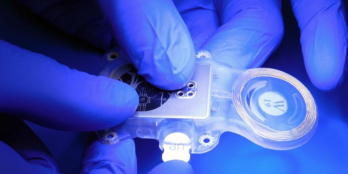
A new study reveals that lignin — a natural plant polymer — can be transformed into a bioactive material that promotes the formation of bone-like minerals while supporting the growth of bone-forming cells. Inspired by the natural partnership between lignin and silica in plants, the research offers a promising step toward sustainable, plant-based materials for future bone regeneration therapies.

A naturally abundant plant material best known for giving trees and crops their strength may one day help repair broken bones, according to a new study led by postdoctoral researcher Dr. Srinath Palakurthy and Prof. Rivka Elbaum of the Hebrew University of Jerusalem. The research demonstrates that lignin — a major structural component of plants — can be engineered into a bioactive material that encourages the formation of hydroxyapatite, the mineral that gives human bones and teeth their strength.

Published in ACS Biomaterials Science & Engineering, the study offers a promising step toward more sustainable, plant-based alternatives to current bone graft materials, many of which are derived from animals or synthetic sources. Such materials are increasingly sought after as researchers work to develop safer, more environmentally friendly solutions for repairing damaged bone.

Structure and evolutionguided design of minimal RNAguided nucleases

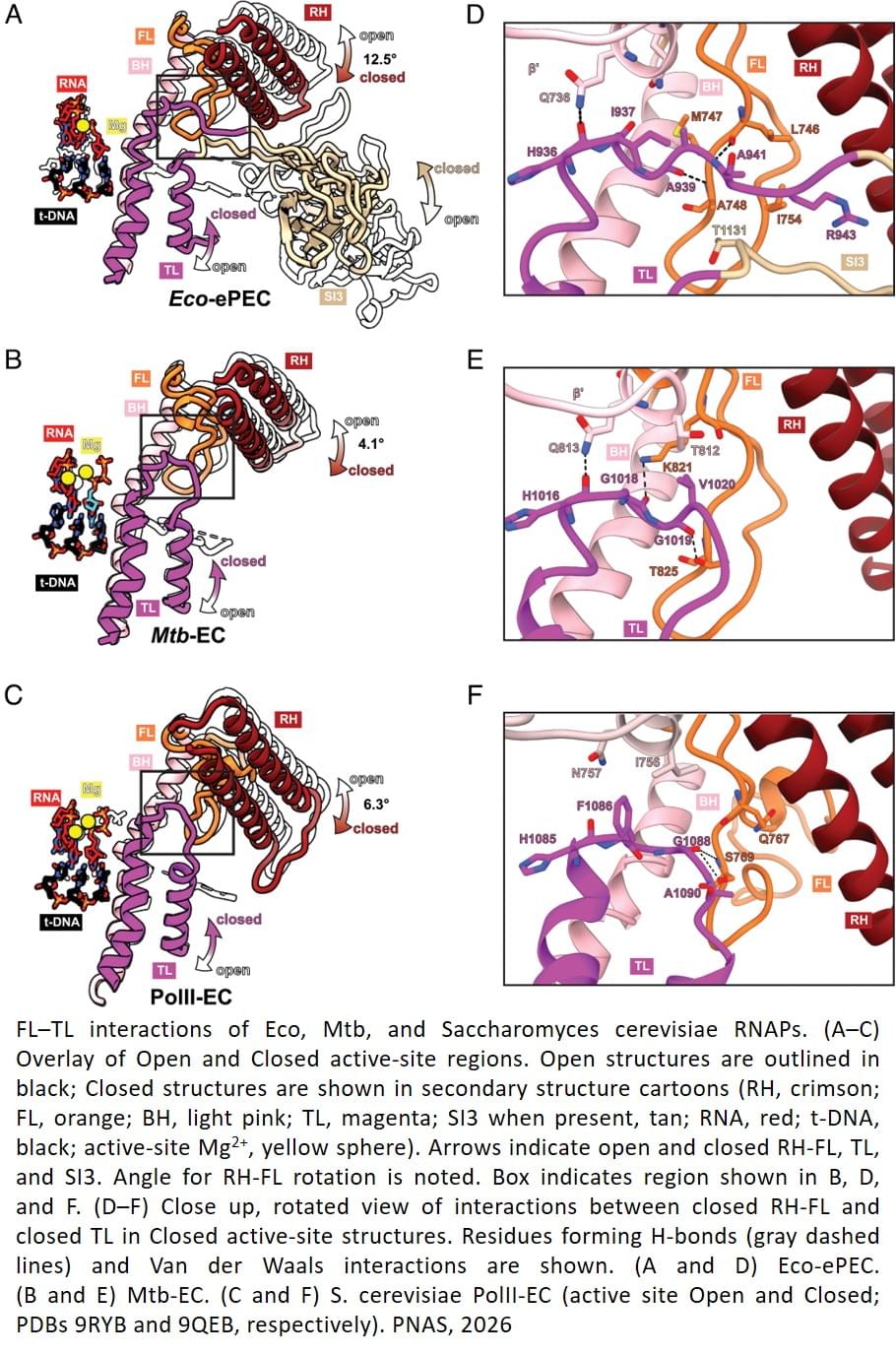
The design of RNA-guided nucleases with properties not limited by evolution can expand programmable genome-editing capabilities. However, generating diverse multidomain proteins with robust enzymatic properties remains challenging. Here, we use a protein design strategy that couples a structure-guided inverse-folding model with evolution-informed residue constraints to generate active, divergent variants of TnpB, a minimal CRISPR-Cas12–like nuclease, termed SynTnpBs. High-throughput screening of artificial intelligence–generated variants yielded editors that retained or exceeded wild-type activity in bacterial, plant, and human cells. Cryo–electron microscopy–based structure determination of the most divergent variant revealed stabilizing contacts in the RNA–DNA interfaces across conformations, demonstrating the design potential of this approach.

Can ctDNA Predict Both Benefit and Toxicity From Immunotherapy?

The investigators asked whether ctDNA status could simultaneously identify patients with a lower probability of therapeutic benefit and a higher likelihood of developing immune-related toxicity. If confirmed, ctDNA could become an important tool not only for predicting efficacy but also for improving treatment selection and avoiding unnecessary exposure to checkpoint inhibitors.

Study Design

The investigators performed a post hoc biomarker analysis of the randomized phase III IMvigor010 trial.

Let’s talk about my clone…

He makes sure to tell people he did not make embryos.


Friends, I did NOT create a human clone. What I did create are induced pluripotent stem cells (iPSCs) — cells that can potentially become many different cell types and may one day help repair the body.

It’s one of the most promising technologies in regenerative medicine, and is already being explored in clinical research.

The science is fascinating, the ethical questions are important, and both deserve thoughtful discussion.

https://blueprint.bryanjohnson.com/?&…

The Role of Lysophosphatidic Acid in Neuropsychiatric and Neurodegenerative Disorders

Individuals suffering from diverse neuropsychiatric and neurodegenerative disorders often have comparable symptoms, which may underline the implication of shared hereditary influences and the same biological processes. Lysophosphatidic acid (LPA) is a bioactive phospholipid and a crucial regulator of the development of adult neuronal systems; hence, it may play an important role in the onset of certain diseases such as Alzheimer’s, Parkinson’s disease, and schizophrenia. During development, LPA signaling regulates many cellular processes such as proliferation, survival, migration, differentiation, cytoskeleton reorganization, and DNA synthesis. So far, six lysophosphatidic acid receptors that respond to LPA have been discovered and categorized based on their homology.

Dreams drain energy: The REM sleep paradox

The brain demands a lot of energy compared with other organs. However, it can also make do when energy supplies are scarce, flexibly processing information using what is available. How the brain resourcefully allocates this limited energy across internal states remains a key question in neuroscience.

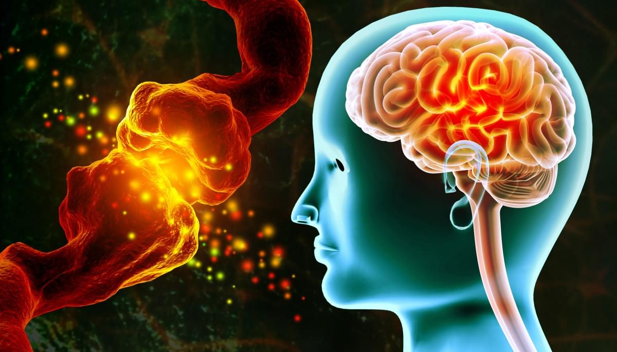
Sleep provides a useful window into answering this question. Although sleep is associated with rest, the brain remains highly active. This is especially true during rapid eye movement (REM) sleep, the stage closely linked to dreaming and memory processing. REM sleep is sometimes called “paradoxical sleep” because the body is largely still while the brain shows wake-like activity. Researchers at Tohoku University have now uncovered another paradox within REM sleep: While energy supply to the dreaming brain appears to rise, the energy molecule used directly by neurons falls. The findings are published in Communications Biology.

“Ever felt exhausted after a vivid dream?” asks Professor Ko Matsui of Tohoku University. “Sleep may appear peaceful, but the brain is highly active—especially when dreaming. We were intrigued by this paradox and wanted to look into the scientific basis behind why dreaming is somehow tiring.”

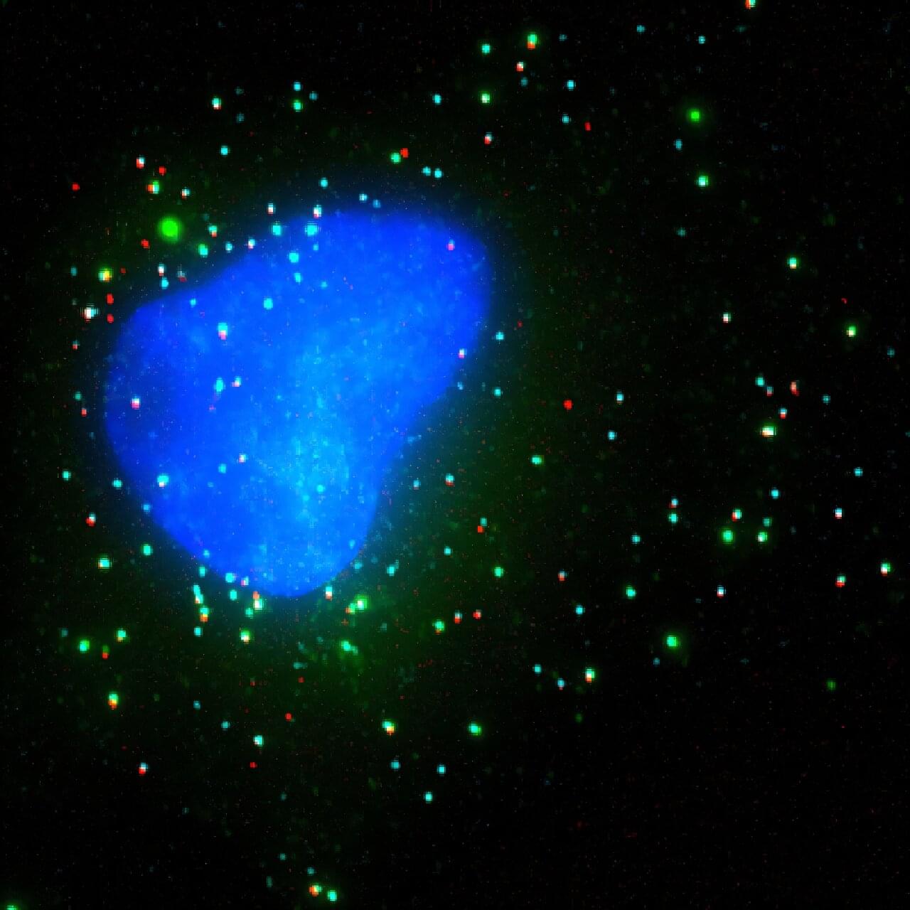
Scientists decipher how T cells sense enemies—such as cancer—at point of contact

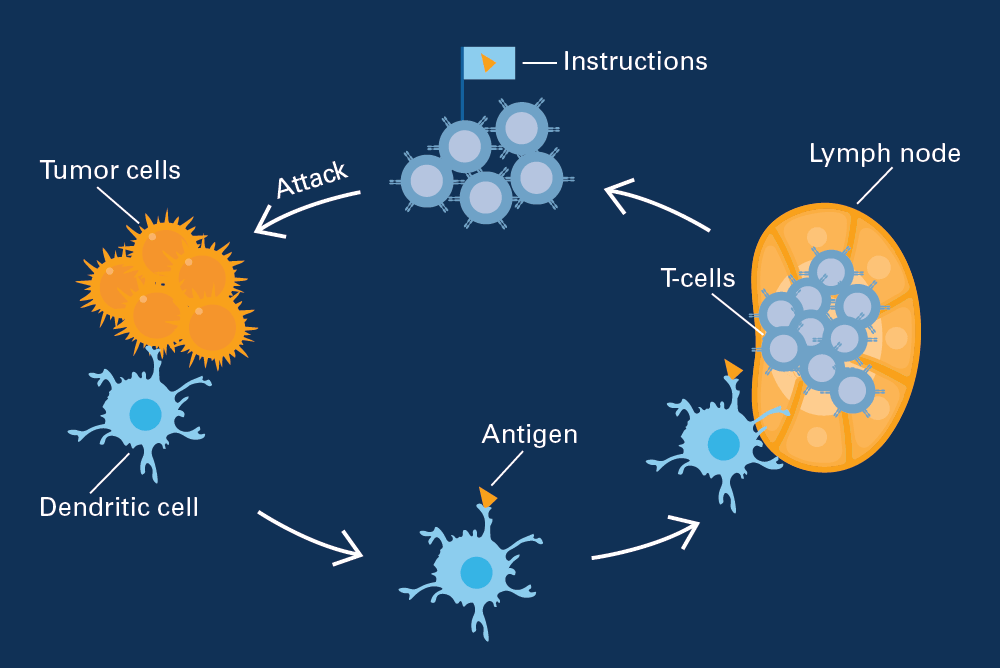
Every encounter between a T cell and a potential target—especially when that target is a developing tumor—begins with a rapid series of molecular decisions. Within seconds, the immune cell must determine whether to launch an attack or stand down. T cells are so potent, so potentially devastating, that misreading the situation can cause serious tissue injury.

But cancer cells come equipped with a bag of tricks that allows them to disarm these powerful warriors of the immune system. All of these activities, whether mediated by T cells or their targets, occur at split-second speed and unfold at the point of cell-to-cell contact.

Now, scientists have identified tiny nanoscale contact points where those decisions are made, revealing how activation and inhibitory signals are integrated at the first moments of a cell-to-cell encounter.

/* */