Transient forebrain ischemia is associated with selective neuronal vulnerability and persistent memory deficit. This study compares functional outcome and morphological changes in rats subjected to post-ischemic CA1 or hilus/dentate gyrus region hippocampal fetal transplantation. Ischemia was produced by bilateral common carotid artery occlusion with hypotension. Fetal hippocampal neurons were transplanted into both sides of the CA1 or hilus/dentate gyrus region of the dorsal hippocampus, 1 week post-ischemia. Four weeks post transplantation, the rats underwent behavioral testing for 5 consecutive days using the water maze trial. All animals were perfusion fixed for morphological studies. Transplants in the CA1 region of the dorsal hippocampus were associated with memory and morphological recovery, while grafts placed into the hilus/dentate gyrus region of the dorsal hippocampus were not. Similarly, neurons transplanted in the CA1 region of the dorsal hippocampus were morphologically similar to CA1 pyramidal cell neurons and stained positive with calbindin D(28k). In contrast the grafts transplanted into the hilus/dentate gyrus region of the dorsal hippocampus were morphologically heterogeneous and staining with calbindin D(28k) was not as robust. Post-ischemic transplantation in the CA1 region of the dorsal hippocampus is effective in improving memory and morphological function.
Category: biotech/medical – Page 64
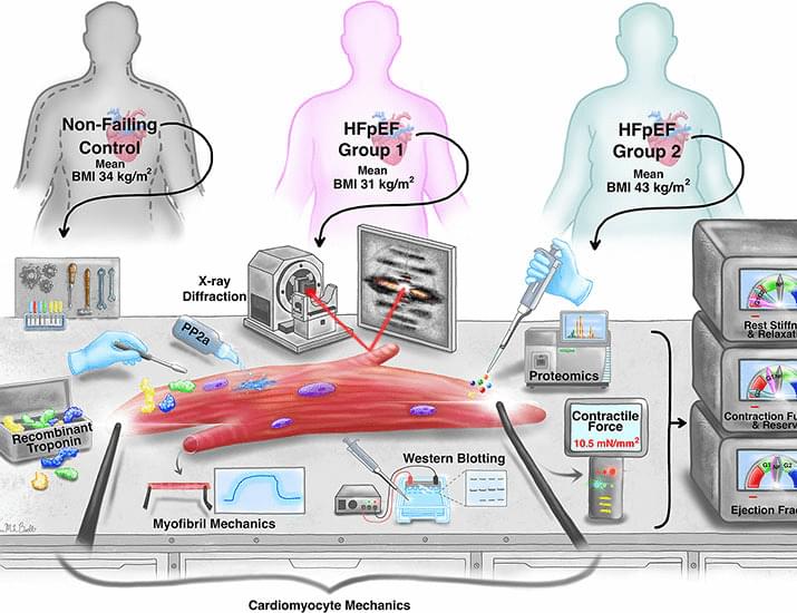
Severe obesity in human HFpEF alters contractile protein function and organization
Heart failure with preserved ejection fraction (HFpEF) causes substantial morbidity and mortality and has few effective therapies. Its phenotype has changed over time, with morbid obesity and metabolic defects supplanting hypertension and cardiac hypertrophy. We reveal that cardiomyocytes from patients with severe obesity and HFpEF have very depressed contractile reserve, including reduced calcium-and length-stimulated tension, power, and myosin activation compared with less-obese HFpEF and nonfailing (NF) controls with or without obesity but similar to those with advanced HF and reduced ejection fraction. Myocyte defects correlate with body mass index and exercise hemodynamics in patients with HFpEF but not NF and appear reversible upon weight loss. Increased troponin I phosphorylation at threonine 181 occurs only in heart failure with obesity, contributing to sarcomere dysfunction.

An ensemble pipeline, PhageHost, for phage tail fiber discovery and accurate Klebsiella pneumoniae host prediction using protein language models
Wu et al. present an ensemble pipeline, PhageHost, comprising a protein language model, TailSeek, for tail fiber detection from phage and prophage genomes and a deep learning model, HostBuster, that integrates tail fiber features with host information to predict the lytic potential of phage–K. pneumoniae pairs.
AI ‘super-antigen’ vaccine could protect against whole families of viruses
A groundbreaking new vaccine technology using artificial intelligence could offer immunity against entire families of viruses and protect against future mutations with a single injection.
Researchers say this could prevent future pandemics before they emerge, saving millions of lives and sparing countries from the necessity of lockdowns.
A “super-antigen” has been developed through AI machine learning that meticulously analyses past and current outbreaks to pinpoint the essential elements for the survival of viruses.
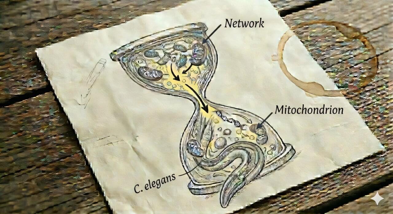
Why energy fades with age: Missing membrane lipid may destabilize mitochondria
Why do cells age—and why do we lose our energy and vitality as we get older? This question is one of the central challenges of modern biomedicine. The focus is particularly on mitochondria—tiny cellular organelles long known as the cell’s powerhouses but now understood as dynamic control centers that not only produce energy, but also coordinate cellular communication, adaptation, and many of the processes essential for life.
They supply us with the energy that our body needs for movement, growth, and repair processes. But as we age, these powerhouses begin to slow down. It has long been known that their function declines with age. But until now, the mechanisms driving this gradual decline have been poorly understood.
Focus on membrane lipids For a long time, it was assumed that genetic damage within the mitochondria themselves was primarily responsible. A study now published in Nature Communications by an international research team led by Dr. Maria Ermolaeva of the Leibniz Institute on Aging—Fritz Lipmann Institute (FLI) in Jena provides a surprising answer to this question: A key factor appears to be the imbalance in the structure of the mitochondrial network, which is caused by the absence of a major lipid in the membrane composition.
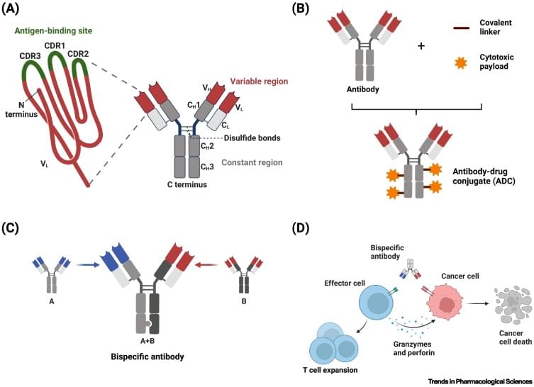
Strategies to boost antibody selectivity in oncology
Antibodies in oncology are being equipped with toxic cargoes and effector functions that can kill cells at very low concentrations. A key challenge is that most targets on cancer cells are also present on at least some healthy cells. Shared targets can result in off-tumor binding and compromise the safety and potential of therapeutic candidates. In this review, we survey strategies that can help direct biologics to cancer sites more selectively. These strategies are becoming increasingly feasible thanks to advances in molecular design and engineering. The objective is to create therapeutics that exploit changes in cancer and leverage the human body infrastructure, enabling therapeutics that discriminate not just self from non-self but diseased from healthy tissue.
The Hidden Impact: Lingering Brain Injury Symptoms Haunt Concussion Patients
Even mild concussion can cause long-lasting effects to the brain, according to researchers at the University of Cambridge. Using data from a Europe-wide study, the team has shown that for almost a half of all people who receive a knock to the head, there are changes in how regions of the brain commu
How cells fight infection from the inside: Newly identified ADX pathway may broaden understanding of immunity
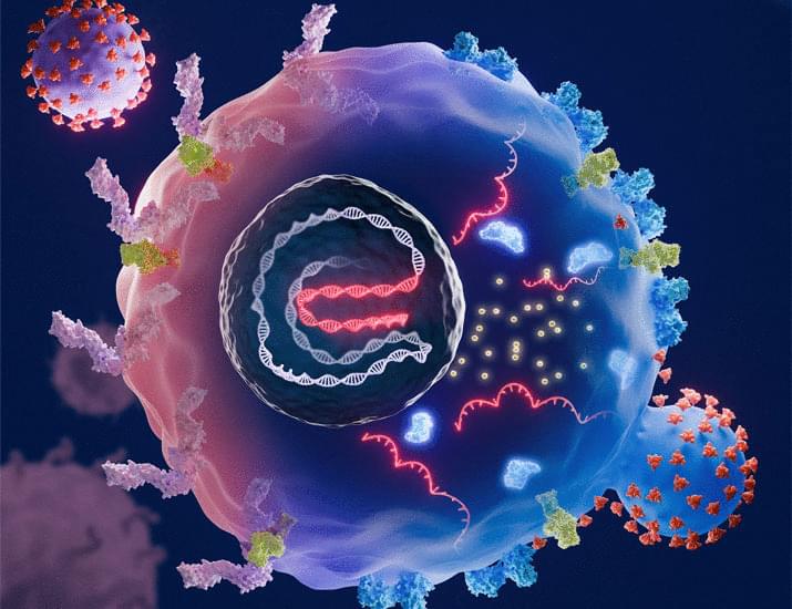
When thinking of the immune system, most people imagine white blood cells putting up a fight against invading germs in the bloodstream. But now, in research published in Molecular Cell, scientists detail a separate but equally important route by which our bodies fight infection—directly inside already infected cells.
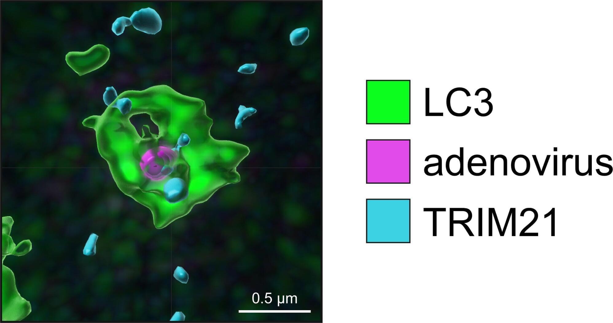
In the report, the authors define a previously undescribed method of germ resistance they coin “antibody-directed xenophagy” (ADX), where cells can digest bacteria and viruses that cross the cell membrane, including Salmonella and adenoviruses.
“People have talked about viral xenophagy before as a sort of concept, but if you look in literature, there aren’t any good examples where people have shown this operating to potently block infection,” says Leo James of the MRC Laboratory of Molecular Biology.
Novel synthetic biomolecule degrades disease-related proteins
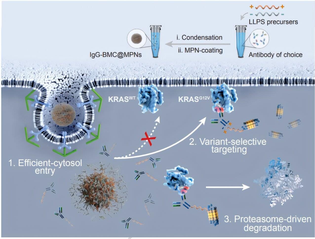
Northwestern Medicine scientists have developed a novel synthetic biomolecular condensate that can degrade intracellular disease-causing proteins, providing a framework for new therapeutic approaches for a wide range of diseases, as detailed in a recent study published in Nature Communications.
Shana Kelley, Ph.D., the Neena B. Schwartz Professor of Chemistry, Biomedical Engineering, and Biochemistry and Molecular Genetics and the president of the Chan Zuckerberg Biohub Chicago, was senior author of the study.
Targeted protein degradation is an emerging therapeutic strategy that harnesses cells’ own degradation machinery to clear disease-causing proteins. However, achieving this degradation process across different cell types has remained a challenge due to subtle variations in protein structure.