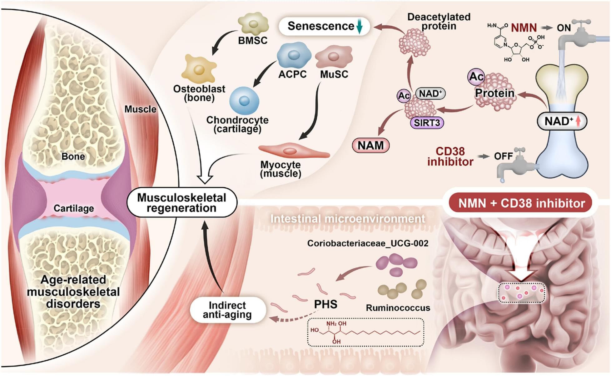
Toggle light / dark theme

Tooth–bone attachment tissue is produced by cells with a mixture of odontoblastic and osteoblastic features in reptiles

Several types of tooth–bone attachment have evolved in different branches of amniotes. The most studied type of tooth anchorage is thecodont implantation, characterized by a nonmineralized periodontal ligament linking the tooth to the jawbone inside a deep alveolus (Bertin et al., 2018 ; Diekwisch, 2001). This attachment, called gomphosis, is present in mammals and crocodilians and provides robust resistance to mechanical stress during food processing (McIntosh et al., 2002).

By contrast, the teeth of recent lepidosaurian reptiles are firmly attached to the jaw bones, although the morphology of this type of attachment varies across species (Gaengler, 2000). In most lizards and snakes, teeth are ankylosed to the inner side of the high labial wall of the jawbone (pleurodont attachment). However, in some species (e.g., agamas, chameleons), the teeth are completely fused to the crest of the tooth-bearing bone (acrodont teeth) (Edmund, 1960). Such cases, where the teeth are firmly fused to the tooth-bearing element by mineralized tissue, are called ankylosis (for nomenclature, see a recent review by Bertin et al., 2018). Although ankylosis is widespread in nature, in mammals, a fusion of the tooth to the bone by hard tissue is considered a pathological condition (Palone et al., 2020 ; Tong et al., 2020).

Diverse developmental mechanisms have been proposed to explain the evolutionary origin and elaboration of ankylosis. The first developmental step of ankylosis is described as a soft ligament mineralization (LeBlanc et al., 2016 ; Liu et al., 2016). The periodontal ligaments in ancestral mammals have been predicted to display a high osteogenic potential, with an inclination to become calcified, thus resulting in dental ankylosis (LeBlanc et al., 2016). The mineralized ligamentous tissue has been preserved in fossilized mosasaurs, and it is also evident in several fish species and modern snakes (LeBlanc, Lamoureux, & Caldwell, 2017 ; Luan et al., 2009 ; Peyer, 1968). In the second type, ankylosis has been described as developing without ligament formation, with the tooth base firmly attached directly to the top of the tooth-bearing bony pedicles with no sign of previous ligament production (Buchtová et al., 2013 ; Luan et al., 2009).

Stem cell organoids mimic aspects of early limb development

Scientists at EPFL have created a scalable 3D organoid model that captures key features of early limb development, revealing how a specialized signaling center shapes both cell identity and tissue organization.

During early development, the embryo builds the body’s organs by exchanging chemical signals between different cell types. When developing limbs, a thin band of skin cells at the limb’s surface, called the “apical ectodermal ridge” (AER), sends signals that guide the underlying population as it grows and forms bone, cartilage, and connective tissue.

The AER is hard to study because it forms only briefly in the embryo and secretes several types of signaling molecules at once. Studying these interactions in embryos is difficult, so scientists often turn to organoids, tiny lab-grown organs that offer researchers a controlled way to study how cells behave and interact as tissues form.

Electrotherapy using injectable nanoparticles offers hope for glioblastoma treatment

Electrotherapy using injectable nanoparticles delivered directly into the tumor could pave the way for new treatment options for glioblastoma, according to a new study from Lund University in Sweden.

Glioblastoma is the most common and most aggressive form of brain tumor among adults. Even with intensive treatment, the average survival period is 15 months. The tumor has a high genetic variation with multiple mutations, which often makes it resistant to radiation therapy, chemotherapy and many targeted drugs. The prognosis for glioblastoma has not improved over the past few decades despite extensive research.

Switching risk and protective alleles improves Alzheimer’s-disease-like signatures and disruptions in mice

Alzheimer’s disease (AD) is a neurodegenerative disorder characterized by the progressive degradation of brain cells, as well as an associated decline in memory and other mental functions. Earlier research found that different forms (i.e., alleles) of a gene known as apolipoprotein E (APOE) are associated with an increased or decreased risk of developing AD.

The APOE gene can be mutated into different variants (i.e., alleles), including APOE2, APOE3 and APOE4. Past studies have linked the presence of two APOE4 alleles to a higher risk of developing AD, while two APOE2 alleles were linked to a significantly lower risk of AD.

Researchers at the University of Kentucky and other institutes genetically engineered a type of mouse that carries a genetic “switch” that can be activated with a drug and that converts the harmful APOE4 allele into the protective APOE2 allele.

Hormone-disrupting chemicals from plastics shown to promote a chronic inflammatory skin condition

A Johns Hopkins Medicine study involving a dozen people with the inflammatory skin disease hidradenitis suppurativa (HS), which mostly affects skin folds, is believed to be the first to provide evidence that hormone-disrupting chemicals commonly found in ultra-processed food and single-use water bottles may contribute to the development of or worsen the condition in some people.

The new findings about the disorder build on previous reports about the role of endocrine-disrupting chemicals, a common environmental contaminant known to mimic, block or alter the body’s hormones, in human health. Researchers believe their findings suggest that reducing exposure could ease HS symptom severity and provide a new avenue of relief for a disease with limited FDA-approved treatment options that include biologic therapy and surgery.

The full report on the study was published in Nature Communications on Nov. 28 and includes insights into the molecular mechanisms that are involved in the disease.

Unexpected pathway for IgA antibody production may help improve vaccines

Scientists led by Stephanie Eisenbarth, MD, Ph.D., the Roy and Elaine Patterson Professor of Medicine and director of the Center for Human Immunobiology, have discovered how critical IgA antibodies are produced through unexpected cellular pathways, findings that may help inform the design of more effective vaccines to prevent infections, according to a recent study published in Immunity.

Immunoglobulin (Ig)A is an antibody that serves as the first line of defense for mucosal tissues that comprise the inner lining of organs in the respiratory system and digestive system. IgA antibodies play a role in humoral immunity, in which IgA and other antibodies produced by B-cells fight off and prevent the spread of infection.

However, inducing an IgA-specific immune response, particularly through vaccines, has remained unsuccessful, according to Eisenbarth.

HIE-ISOLDE: Ten years, ten highlights

The Isotope Separator On-Line facility (ISOLDE) directs a proton beam from the Proton Synchrotron Booster (PSB) onto specially developed thick targets, producing low-energy beams of radioactive nuclei—those with too many or too few neutrons to be stable. These beams can be further accelerated to energies of up to 10 MeV per nucleon using the HIE-ISOLDE linear accelerator, enabling a wide range of studies.

The HIE-ISOLDE beams are sent to three experimental stations: the Miniball array of high-purity germanium gamma-ray detectors, the ISOLDE solenoid spectrometer (ISS), which repurposed a former MRI magnet, and the scattering experimental chamber (SEC), used for a broad variety of physics experiments. Since its first experiment in October 2015, HIE-ISOLDE has been pushing back the boundaries of nuclear physics. To celebrate its 10th anniversary, we look back at 10 key achievements that have defined its first decade.

/* */