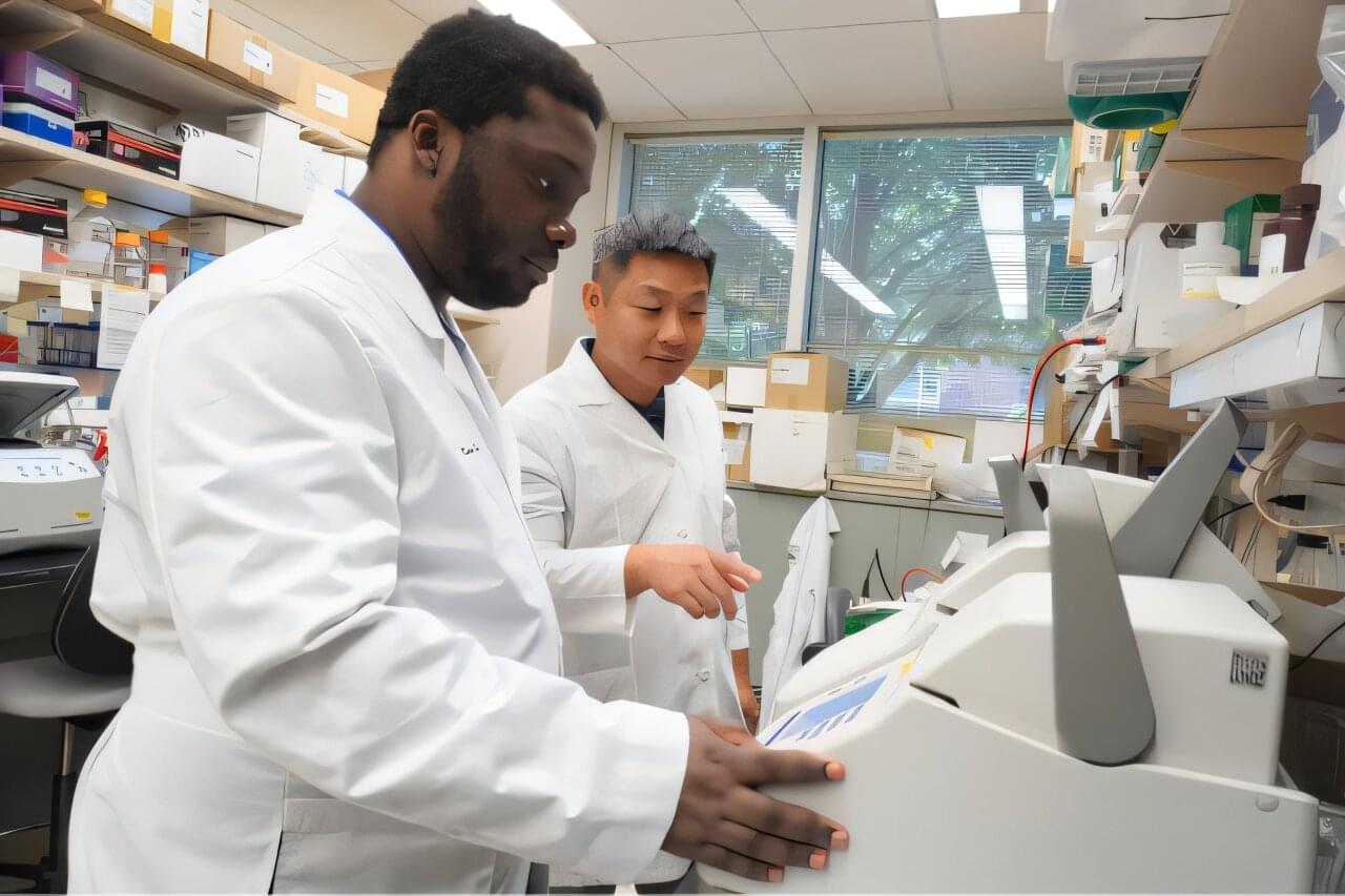
Toggle light / dark theme

Abstract: Expanding clones, expanding aneurysms through macrophage-to-osteoclast differentiation:

Jessica A. Regan & Svati H. Shah Comment on Yonekawa et al.: https://doi.org/10.1172/JCI198708 aneurysm.


Address correspondence to: Jessica A. Regan, Duke Molecular Physiology Institute Duke University School of Medicine, 300 N. Duke Street, Carmichael Building, Durham, North Carolina, 27,701, USA. Email: [email protected].

Real-time impedance-based cell migration measurements with integrated electrodes on porous membranes for next generation microphysiological systems

A new laboratory technique for measuring how quickly cells penetrate and pass through a porous membrane and reach the opposite side could help identify cancer cells with the greatest potential to spread in the human body.

The method relies on tiny electrodes placed on either side of an artificial membrane. The electrodes measure changes in electrical resistance as cells pass through the material. The most aggressive cancer cells pass through the membrane more rapidly than other cells.

The illustration depicts cells (green and blue) moving through a membrane (grey) studded with microelectrodes (gold rings).

🔗


We present a novel microfluidic device capable of electrically interrogating both surfaces of a porous membrane quantitatively and in real time using electrical impedance spectroscopy to monitor cell migration. This device holds patterned gold electrodes on both sides of the membrane, which enable independent impedance measurements on each side of the membrane. We introduce the term cross-over cell migration (CoCM) to describe this dual-sided approach, which allows precise monitoring of cells at their seeding location and as they move through a porous membrane. To ensure reliable tracking, we developed a normalization method, the CoCM index, that allows us to compare both membrane surfaces directly in real-time. Human renal carcinoma cells (786-O) were passively seeded in the device’s top microfluidic chamber, and we collected impedance data from both sides of the membrane surfaces simultaneously over a three-day period. These measurements successfully captured the onset and progression of cell migration across the membrane interface. We tracked the cells with fluorescence imaging in parallel to validate our impedance data. As cells appeared in focus on the bottom-side electrode surface, their numbers kept increasing over the course of our experiment. The CoCM index decreased by about 20% in the top chamber and increased by approximately 15% in the bottom chamber. Symmetrical CoCM index trends appeared after 40 h, consistent with the fluorescent images captured. Finally, we performed live-cell fluorescence assays to confirm post-experiment cell viability and to quantify migrated cells, further validating our CoCM platform measurements. This platform is a valuable tool not only for real-time and quantitative cell migration studies of cancer and other cells in bulk but also for future studies of single-cell migration processes.

This artificial retina doesn’t just aim to restore sight—it opens a hidden channel of vision

The retina, the thin layer of tissue at the back of the eye, is made up of photoreceptor cells that convert visible light into electrical signals, which is essential for human vision. Some diseases, such as retinal degeneration, cause these photoreceptor cells to stop working, which results in blindness. Researchers at Yonsei University, the Institute for Basic Science (IBS) and other institutes in the Republic of Korea have recently developed a new artificial retina that could partly restore vision in people with damaged retinas.

The new device, introduced in a paper published in Nature Electronics, works by detecting near-infrared light and converting it into electrical signals, which stimulate another type of cells in the retina that are undamaged.

“Many people suffer from blindness due to retinal diseases that cause photoreceptor degeneration,” wrote Won Gi Chung, Inhea Jeong and their colleagues in their paper. “Electrical stimulation of retinal neurons can recreate the action potentials associated with seeing that are generated by these cells. We report a thin artificial retina that can be adhered to the epiretinal surface and can convert near-infrared (NIR) light into electrical stimuli that selectively stimulate ganglion cells.”

Subversion of kynurenine-induced AHR activation in CD8 T cells by kynureninase-expressing antigen-presenting cells

Giacomantonio et al. demonstrate an immunomodulatory role for a metabolic enzyme kynureninase in antigen-presenting cells (APCs). Kynureninase-expressing APCs degrade kynurenine, including that generated by cancer cells, and relieve kynurenine-induced immunosuppression in CD8+ T cells.

Gene therapy for deafness approved

The world’s first gene therapy for deafness received approval from the U.S. Food and Drug Administration today. The treatment, from biotech company Regeneron, targets hearing loss caused by inherited mutations in the OTOF gene, which encodes otoferlin, a protein that allows the inner ear’s hair cells to sense and transmit sound to the brain. Patients receive a one-time ear injection containing viral vectors that carry a working copy of the OTOF gene into their cells. In a clinical trial, nine of 12 deaf children who initially received the Regeneron therapy gained enough hearing to stop using cochlear implants; three within that group ended up having normal hearing. Although many gene therapies cost $1 million or more, Regeneron said its treatment, called Otarmeni, will be free in the United States.

Eli Lilly & Co. and researchers in China are also developing gene therapies for OTOF mutations, which account for up to 3% of cases of inherited deafness. One U.S.-Chinese team reported in Nature this week that among 24 patients, including some adults, hearing improvements have lasted more than 2 years in some cases, NPR reports. Researchers eventually hope to treat other types of genetic deafness as well, but those attempts face more challenges. For example, for some disorders, it may be necessary to regenerate lost hair cells. In others, targeting the wrong cell type could damage hearing.

Research Into Naturally Occurring Hair Growth in Skin Nevi May Inform New Regenerative Therapies

An international team of researchers funded in part by NIAMS sought to understand why skin nevi grow long hair. Nevi, which are a type of skin lesion, have an abundance of pigment-producing cells, called melanocytes, that have become aged (or senescent). The team determined that senescent melanocytes within nevi produce large quantities of several signaling molecules. One such molecule, called osteopontin, causes dormant hair stem cells to wake up, which increases hair growth.

The study, which appeared in the journal Nature on June 21, 2023, provides answers as to why nevi are hairy and also uncovers the unexpected growth-promoting potential of senescent cells, which are typically thought to be associated with inhibited tissue growth.

Identification and characterization of BRAF⇔TP53 interactions in melanoma

O’Toole et al. identify novel interactors of both normal BRAF and BRAFV600E and discover TP53 as a BRAFV600E-enhanced interactor in melanoma cells. While TP53 mutations do not frequently occur in melanoma, the authors demonstrate TP53 inactivation and a sequestration of cytoplasmic TP53 after oncogenic BRAF activation.

Recent Scientific Evidence that Supports Nichols’s Lost Primal Eye Theory of Mind I. Core Premise: The Evolutionary Shift

The Phantom Organ and The “Hard Problem” — I apply MVT to solve David Chalmers’s “Hard Problem” of consciousness-the question of why physical brain processes are accompanied by subjective feelings (qualia).


Nichols’s theory posits that self-referential consciousness and abstract thought in many modern animals are the evolutionary result of the loss of a physical sensory organ: the parietal/pineal eye (the “primal eye”). Nichols maps this transition across three brain states in vertebrate evolution: The E2 State (Finite-State): Early fish, amphibians, and ancestral reptiles (as well as modern “living fossils” like the Tuatara) possessed a functional, light-sensitive median eye on top of their skulls, connected to the pineal gland. This organ directly controlled thermoregulation, circadian rhythms, and basic predator detection in coldblooded (ectothermic) animals. Their brains were “hard-wired,” responding directly to environmental stimuli. The E1 State (Infinite-State): As mammals and birds evolved warmbloodedness (endothermy), external temperature sensing became redundant, and advanced lateral eyes took over visual duties. The primal eye atrophied, leaving behind only the internal pineal gland. Freed from the direct “lock-step” control of the sun, the brain became plastic and self-organising (infinite-state). The E0 State: Some lineages, like certain dinosaurs and modern crocodilians, lost both the median eye and the pineal gland entirely. II. The Phantom Organ and The “Hard Problem” Nichols applies MVT to solve David Chalmers’s “Hard Problem” of consciousness-the question of why physical brain processes are accompanied by subjective feelings (qualia). The Virtual Sensor: Just as an amputee can experience a “phantom limb” because the neural matrix still expects the arm, the E1 mammalian brain experiences a “phantom eye”. The brain was built over millions of years to process a central stream of generic sensory data from the primal eye. Centrally Evoked Mentation: When the physical eye retreated, it left an internal sensory void. The brain compensated by simulating the presence of this lost hub to unscramble data from the other senses. This virtual simulation is the seat of the subjective “I”. III. The Origins of REM Sleep and Dreaming Nichols heavily critiques philosophers like Owen Flanagan, who argue that dreams are useless evolutionary “spandrels” (biological noise). Baseline Architecture: In MVT, Rapid Eye Movement (REM) sleep is the baseline functional state of the new E1 architecture. Because the physical tether to sunlight was severed, the brain uses this “phantom” space to generate internal models.

New cellular target prevents hepatitis E infection

An international team of researchers has identified a promising new approach for treating infections with the Hepatitis E virus (HEV). At the center of the study is the drug Apilimod, which specifically blocks the entry of the virus into human liver cells, thereby preventing infection at an early stage. The compound targets a mechanism of the host cell, reducing the likelihood that the virus will develop resistance.

Apilimod has already been clinically evaluated, which could accelerate its development into a drug against hepatitis E. The study, led by the Department of Molecular and Medical Virology at Ruhr University Bochum, Germany, was published in the journal eGastroenterology on March 31, 2026.

Autoantibody map uncovers body-wide immune attacks across Alzheimer’s, Parkinson’s and MS

Researchers at the University of São Paulo (USP) in Brazil discovered that neurodegenerative diseases, such as Alzheimer’s, Parkinson’s, and multiple sclerosis, are more complex than previously thought. Their analysis of nearly 600 blood samples from patients with and without these diseases revealed that neurodegenerative processes extend beyond the central nervous system, affecting various targets throughout the body.

“We conducted a systemic analysis based on autoantibodies—defense proteins [immunoglobulins] that mistakenly attack the body’s healthy cells, tissues, or organs instead of external pathogens. In this study, we saw that, contrary to what was previously thought, these diseases don’t involve an antibody attacking only a specific region of the connection between neurons [synapse], like a thief breaking in through a door. It’s a systemic attack, like machine-gunning an entire house,” explains JĂșlia Nakanishi Usuda, first author of the study.

The study, published in the journal iScience, identified more than 9,000 autoantibodies from public databases. Based on the results, the researchers suggest that, rather than focusing on isolated molecular targets, treatment strategies for these diseases should focus on blocking the autoimmune response systemically. While the data science study still needs to be confirmed through in vitro and in vivo testing, it reinforces a new paradigm for treating neurodegenerative diseases.

/* */