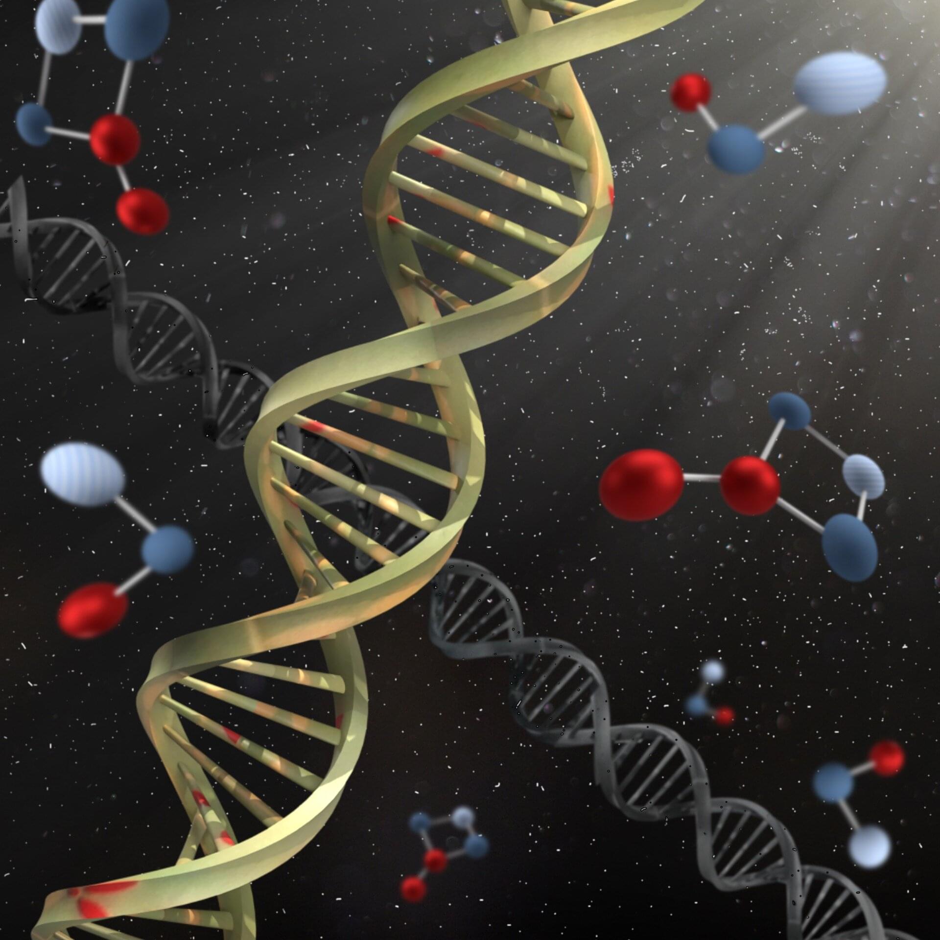
Toggle light / dark theme

Why no individual is like another when epigenetics come into play

Why do animals behave differently, and what are the consequences of this? A research team from the Collaborative Research Center NC³ at Bielefeld University and the University of Münster now provides a new explanation: epigenetic processes—chemical markings on DNA—may play a key role. The study, published in the journal Trends in Ecology & Evolution, links individuality, environmental adaptation, genetics, ecology, and evolution in a novel way.

“With our study, we propose that individuality and epigenetic variation influence each other,” explains Dr. Denis Meuthen, an evolutionary biologist at Bielefeld University, who is one of the study’s main authors. “This bidirectionality—this mutual interaction—helps us to better understand ecological and evolutionary processes.”

RNA-guided CRISPR system activates gene expression

In back-to-back studies published in Nature, researchers from Purdue University and Columbia University report a naturally evolved gene-editing system that can activate genes, offering an advantage over existing CRISPR gene-editing systems that merely find and cut DNA. The research includes two complementary studies, one examining the biological function of the system and the other revealing the molecular mechanism that enables it.

The team’s research on a variant of the CRISPR—Clustered Regularly Interspaced Short Palindromic Repeats—system broadens understanding of CRISPR’s natural diversity and provides a foundation for new gene-regulation technologies. Because this CRISPR variant activates genes without cutting DNA, it could be adapted for precise gene control applications, including research tools and potential therapeutic strategies that turn on genes without permanently altering the genome.

One study shows that this CRISPR system, using a strand of RNA as a guide, finds specific sections of DNA, known as genes, and attracts the cell’s own gene expression machinery to the location to activate the gene. The second study explains how the molecular complex performs this task, revealing how its structure allows it to recruit RNA polymerase—the enzyme responsible for transcribing DNA into RNA—to initiate gene expression.

Protein modification discovery opens cancer therapy possibilities

A research team led by Purdue University’s W. Andy Tao has discovered a new type of protein modification related to cellular mutation that impairs a crucial enzyme’s ability to help drive energy processes. Their discovery, published in Nature Chemistry, opens a new route to therapeutic cancer intervention.

“Mutation is considered the driving mechanism leading to cancer. Many mutations are hidden and harmless, but the mutation of enzymes like kinases can lead to the uncontrolled growth of cancer cells,” said Tao, a professor of biochemistry in Purdue’s College of Agriculture.

The study wades into the interactive dynamic complexity of the human genome (containing 20,000 to 25,000 genes) and the human proteome (containing more than 1 million proteins). The researchers identified a new modification on proteins because of the mutation in the isocitrate dehydrogenase (IDH) enzyme, which affects how kinase enzymes control protein function.

Glioblastoma Growth Mechanism Identified, Pointing to Potential Therapeutic Targets

Ruhi Polara, PhD, who led the research alongside Robinson, further commented, “Essentially, CD47 is shielding ROBO2, allowing it to accumulate and drive tumor progression. When we remove CD47, ROBO2 is degraded, and the cancer cells lose their ability to grow and invade effectively.”

The findings reveal a previously unknown molecular pathway—CD47–ITCH–ROBO2—that controls how glioblastoma cells behave. This opens up new possibilities for treatment strategies that go beyond current approaches. While therapies targeting CD47 are already being tested in clinical trials for other cancers, they have shown limited success in glioblastoma so far. The new research suggests that directly targeting the CD47–ROBO2 pathway, or disrupting the stabilisation of ROBO2, could be a more effective strategy. “In summary, our study reveals a role of CD47 in regulating cellular plasticity suggesting that targeting ROBO2 could offer a promising alternative therapeutic strategy for GBM,” they stated.

“By understanding this mechanism, we now have new targets to explore,” Polara said. “This could lead to the development of therapies that specifically block the tumor’s ability to spread, which is one of the biggest challenges in treating glioblastoma.”

Local gene editing of fibroblasts in tumors reveals a new cancer-associated fibroblast state

Nicholas F. Kuhn, Matthew F. Krummel et al. demonstrate how local gene editing of cancer-associated fibroblasts alters their cell state and, subsequently, the cellular tumor microenvironment.


CAFs are prominent members of the TME. Kuhn et al. demonstrate how local gene editing of CAFs alters their cell state and, subsequently, the cellular TME.

FAK inhibition in ovarian cancer releases omega-3 fatty acids to program CXCL13-producing anti-tumor resident peritoneal macrophages

FAK tyrosine kinase drives ovarian cancer tumor progression in part via effects on the tumor microenvironment. Chen et al. show that ovarian tumor FAK inhibition triggers release of omega-3 fatty acid-containing exosomes, impacting GATA6+ peritoneal macrophage anti-tumor reprogramming, CXCL13 cytokine production, and anti-TIGIT immunotherapy.

Monocytes Defined by Platelet Interactions and Oxidative Stress Signaling Underlie HIV‐Associated Atherosclerosis

This study reveals an atherosclerosis-associated signature in platelet-monocyte complexes from people living with HIV. @RuoqiaoW @ThakarLab @URochester_SMD


BackgroundMonocytes contribute to atherosclerosis by migrating into inflamed endothelium and differentiating into lipid‐laden macrophages. In people living with HIV, chronic inflammation increases atherosclerosis risk, yet the role of specific monocyte subsets remains unclear. We investigated how distinct monocyte populations contribute to vascular pathology in early HIV‐associated atherosclerosis.

Early Clinical and EEG Association of Genotype and Outcome in Genetic EpilepsiesA Cohort Study and Hierarchical Clustering Analysis

This study analyzed a large cohort of patients with genetic epilepsies using hierarchical clustering analysis to identify homogeneous subgroups defined by specific genetic causes, each showing distinct clinical and EEG patterns.


We included 277 patients (52.3% female; median age at last follow-up 8.1 years, range 0–40). Drug resistance occurred in 58.8% and severe DD/ID in 35.4% of patients. EEG data at onset were available for 107 individuals. Neonatal onset was associated with a higher rate of drug resistance (71.4%; odds ratio [OR] 2.0, 95% CI 1.05–3.77), movement disorders (60.7%; OR 3.7, 95% CI 2.02–6.82), and severe DD/ID (71.4%; OR 7.0, 95% CI 3.66–13.49). Slow EEG background activity and multifocal epileptiform discharges were associated with both drug resistance and severe DD/ID. HCA identified genotype-phenotype groupings, including clusters involving SCN1A, PRRT2, STXBP1, KCNQ2, SCN2A, CHD2, SYNGAP1, and MECP2, each linked to specific clinical and EEG features.

Single-cell maps show chemokine signals collapse as aggressive lymphoma spreads

Lymph nodes are key control centers in the immune system and play an important role in defending the body against infections and tumors. For these processes to function properly, immune cells (B cells and T cells) must be organized in a precise spatial pattern in the lymph node tissue, for example in so-called B cell follicles and T cell zones. They are controlled by stromal cells (non-hematopoietic structural cells). They release messenger substances called chemokines, creating signals to guide the immune cells to their designated positions in the lymph node.

In the case of B cell lymphomas, the internal structure of the lymph node tissue can be disturbed in very different ways, depending on the exact type of lymphoma: While the fundamental tissue structure remains intact in the case of slow-growing lymphomas such as follicular lymphoma (FL), aggressive lymphomas such as diffuse large B cell lymphoma (DLBCL) cause the tissue structure to break down completely. Why these typical growth patterns develop has been largely unclear to date.

In the study “Architectural principles of lymphoma-induced lymph node tissue remodeling,” the researchers coordinated by Professor Dietrich (Director of the Department of Hematology, Oncology and Clinical Immunology, UKD) have now succeeded in systematically mapping these processes in the human lymph node for the first time. By means of single-cell analyses and spatial tissue mapping, they were able to trace which factors lead to the progressive breakdown of the lymph node architecture in the case of lymphoma. The work is published in the journal Nature Cancer.

/* */