Toggle light / dark theme

Regorafenib.


For patients with metastatic esophagogastric adenocarcinoma, inhibition of the vascular endothelial growth factor (VEGF) pathway via the anti–VEGFR-2 antibody ramucirumab increases response, progression-free survival (PFS) and overall survival (OS) when combined with second-line treatment with paclitaxel. The multitargeted tyrosine kinase inhibitor regorafenib targets VEGFR and other pathways and has effectiveness in chemotherapy-refractory colorectal cancer.

Investigators now report results of the industry-sponsored, randomized, double-blind, placebo-controlled INTEGRATE IIa trial evaluating regorafenib (160 mg daily for 21 days of 28-day cycles) in patients with chemotherapy-refractory esophagogastric adenocarcinoma. Of the 251 patients enrolled, 63% were treated in Asia; 73% had gastric primaries; 42% had received a prior VEGF inhibitor; 59% had received two and 40% had received three or more lines of prior chemotherapy. For the analysis of OS, the primary endpoint, results were pooled with those from INTEGRATE I, a prior randomized, placebo-controlled, phase II trial in 147 patients.

At a median follow-up of 48 months, OS was improved with regorafenib over placebo in the pooled cohort (hazard ratio, 0.70; P=0.001) and in the INTEGRATE IIa cohort (HR, 0.68; P=0.006). In the INTEGRATE IIa cohort, 1-year OS was improved with regorafenib (19% vs. 6%), whereas median OS was similar (4.0 and 4.5 months). PFS was improved with regorafenib (HR, 0.53; P0.0001). The disease control rate was higher with regorafenib than placebo (21.3% vs. 4.9%) and the antitumor response was limited (2.4% vs. 0%). Adverse events related to regorafenib, including palmar–plantar erythrodysesthesia, mucositis, hypertension, and diarrhea, were manageable.

Mapping the geometry of quantum worlds: measuring the quantum geometric tensor in solids.

Quantum states are like complex shapes in a hidden world, and understanding their geometry is key to unlocking the mysteries of modern physics. One of the most important tools for studying this geometry is the quantum geometric tensor (QGT). This mathematical object reveals how quantum states “curve” and interact, shaping phenomena ranging from exotic materials to groundbreaking technologies.

The QGT has two parts, each with distinct significance:

1. The Berry curvature (the imaginary part): This governs topological phenomena, such as unusual electrical and magnetic behaviors in advanced materials.

2. The quantum metric (the real part): Recently gaining attention, this influences surprising effects like flat-band superfluidity, quantum Landau levels, and even the nonlinear Hall effect.

The most common form – obstructive sleep apnoea – happens when the walls of the throat relax and narrow or close, with symptoms including choking noises, loud snoring and waking up a lot.

The three-hour procedure to fit Nyxoah’s Genio implant was carried out by medics at University College London Hospitals NHS foundation trust (UCLH) this month.

One of the two patients, Natalie Boller, 63, was feeling better within days and will return to the clinic to have the device activated in the coming weeks.

Research utilizing AI tool AlphaFold has revealed a new protein complex that initiates the fertilization process between sperm and egg, shedding light on the molecular interactions essential for successful fertilization.

Genetic research has uncovered many proteins involved in the initial contact between sperm and egg. However, direct proof of how these proteins bind or form complexes to enable fertilization remained unclear. Now, Andrea Pauli’s lab at the IMP, working with international collaborators, has combined AI-driven structural predictions with experimental evidence to reveal a key fertilization complex. Their findings, based on studies in zebrafish, mice, and human cells, were published in the journal Cell.

Fertilization is the first step in forming an embryo, starting with the sperm’s journey toward the egg, guided by chemical signals. When the sperm reaches the egg, it binds to the egg’s surface through specific protein interactions. This binding readies their membranes to merge, allowing their genetic material to combine and create a zygote—a single cell that will eventually develop into a new organism.

Join us on Patreon! https://www.patreon.com/MichaelLustgartenPhD

Discount Links/Affiliates:
Blood testing (where I get the majority of my labs): https://www.ultalabtests.com/partners/michaellustgarten.

At-Home Metabolomics: https://www.iollo.com?ref=michael-lustgarten.
Use Code: CONQUERAGING At Checkout.

Clearly Filtered Water Filter: https://get.aspr.app/SHoPY

PHOENIX — Mayo Clinic announces the results of an innovative treatment approach that may offer improvement in overall survival in older patients with newly diagnosed glioblastoma while maintaining quality of life. Glioblastoma is the most lethal type of primary brain cancer due to its aggressive nature and its treatment-resistant characteristics. It is the most common form of primary brain cancer. Each year an estimated 14,500 people in the U.S. are diagnosed with the disease. Results of Mayo Clinic’s phase 2, single-arm study are published in The Lancet Oncology.

Sujay Vora, M.D., radiation oncologist at Mayo Clinic, led a team of researchers investigating the use of short-course hypofractionated proton beam therapy incorporating advanced imaging techniques in patients over the age of 65 with newly diagnosed World Health Organization (WHO) grade 4, malignant glioblastoma.

Results showed that 56% of participants were alive after 12 months and the median overall survival was 13.1 months.” As compared to prior phase 3 studies in an older population having a median survival of only six to nine months, these results are promising,” says Dr. Vora. “In some cases, patients with tumors that have favorable genetics lived even longer, with a median survival of 22 months. We are very excited about these results.”

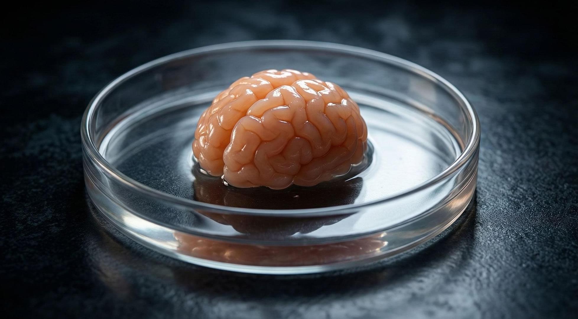
Microgravity is known to alter the muscles, bones, the immune system and cognition, but little is known about its specific impact on the brain. To discover how brain cells respond to microgravity, Scripps Research scientists, in collaboration with the New York Stem Cell Foundation, sent tiny clumps of stem-cell derived brain cells called “organoids” to the International Space Station (ISS).

Surprisingly, the organoids were still healthy when they returned from orbit a month later, but the cells had matured faster compared to identical organoids grown on Earth—they were closer to becoming adult neurons and were beginning to show signs of specialization. The results, which could shed light on potential neurological effects of space travel, appear in Stem Cells Translational Medicine.

“The fact that these cells survived in space was a big surprise,” says co-senior author Jeanne Loring, Ph.D., professor emeritus in the Department of Molecular Medicine and founding director of the Center for Regenerative Medicine at Scripps Research. “This lays the groundwork for future experiments in space, in which we can include other parts of the brain that are affected by neurodegenerative disease.”

Pain is meant to be a defense mechanism. It creates a strong sensation to get us to respond to a stimulus and prevent ourselves from further harm. But, sometimes injuries, nerve damage, or infections can cause long-lasting, severe bouts of pain that can make daily life unbearable.

What if there was a way to simply turn off ? UNC School of Medicine researchers Bryan L. Roth, MD, Ph.D., the Michael Hooker Distinguished Professor of Pharmacology, and Grégory Scherrer, PharmD, Ph.D., associate professor of cell biology and physiology and the UNC Neuroscience Center, have just proven that it is possible.

Using a tool designed by Roth in the early 2000s, the labs have created a new system that reduces acute and tissue-injury-induced inflammatory in mouse models. Hye Jin Kang, Ph.D., an alumnus of the Roth Lab and now associate professor at Yonsei University in Korea, was first author on the research paper. The results were published in Cell.

DDW Editor Reece Armstrong speaks to Ellie Mahjubi, Vice President, Protein and cell analysis at Thermo Fisher Scientific, about how spatial biology is impacting drug discovery and development research.

RA: What’s the future and potential for spatial biology?

EM: Technological advancements in spatial biology are providing unprecedented insights into single cells within their spatial context, facilitating the analysis of cell types, functional states, cell interaction networks, as well as tissue microenvironments and architecture. These innovations promise to significantly advance basic research and enhance our understanding of human health and disease.

Researchers have used a chemical compound to light up treatment-resistant cancers on imaging scans, in a breakthrough that could help medical professionals better target and treat cancer.

The authors at King’s College London say that using the radiotracer—an injected compound used in PET scans—could help inform doctors that a patient’s aggressive cancer will not respond to chemotherapy before treatment is given. This would prevent patients receiving unnecessary treatment and provide them with alternative options that will give them the best chance of beating the disease.

The paper, “Imaging NRF2 activation in non-small cell lung cancer with published in Nature Communications, shows therapy-resistant tumors “lit up like a Christmas tree” on PET scans when the radiotracer was injected.