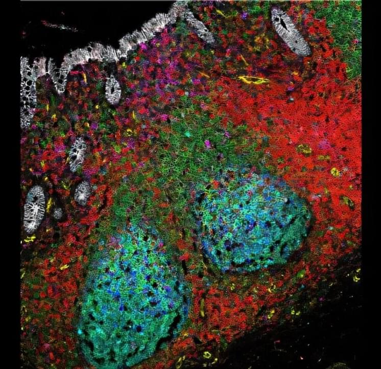
Toggle light / dark theme

Recent Scientific Evidence that Supports Nichols’s Lost Primal Eye Theory of Mind I. Core Premise: The Evolutionary Shift

The Phantom Organ and The “Hard Problem” — I apply MVT to solve David Chalmers’s “Hard Problem” of consciousness-the question of why physical brain processes are accompanied by subjective feelings (qualia).


Nichols’s theory posits that self-referential consciousness and abstract thought in many modern animals are the evolutionary result of the loss of a physical sensory organ: the parietal/pineal eye (the “primal eye”). Nichols maps this transition across three brain states in vertebrate evolution: The E2 State (Finite-State): Early fish, amphibians, and ancestral reptiles (as well as modern “living fossils” like the Tuatara) possessed a functional, light-sensitive median eye on top of their skulls, connected to the pineal gland. This organ directly controlled thermoregulation, circadian rhythms, and basic predator detection in coldblooded (ectothermic) animals. Their brains were “hard-wired,” responding directly to environmental stimuli. The E1 State (Infinite-State): As mammals and birds evolved warmbloodedness (endothermy), external temperature sensing became redundant, and advanced lateral eyes took over visual duties. The primal eye atrophied, leaving behind only the internal pineal gland. Freed from the direct “lock-step” control of the sun, the brain became plastic and self-organising (infinite-state). The E0 State: Some lineages, like certain dinosaurs and modern crocodilians, lost both the median eye and the pineal gland entirely. II. The Phantom Organ and The “Hard Problem” Nichols applies MVT to solve David Chalmers’s “Hard Problem” of consciousness-the question of why physical brain processes are accompanied by subjective feelings (qualia). The Virtual Sensor: Just as an amputee can experience a “phantom limb” because the neural matrix still expects the arm, the E1 mammalian brain experiences a “phantom eye”. The brain was built over millions of years to process a central stream of generic sensory data from the primal eye. Centrally Evoked Mentation: When the physical eye retreated, it left an internal sensory void. The brain compensated by simulating the presence of this lost hub to unscramble data from the other senses. This virtual simulation is the seat of the subjective “I”. III. The Origins of REM Sleep and Dreaming Nichols heavily critiques philosophers like Owen Flanagan, who argue that dreams are useless evolutionary “spandrels” (biological noise). Baseline Architecture: In MVT, Rapid Eye Movement (REM) sleep is the baseline functional state of the new E1 architecture. Because the physical tether to sunlight was severed, the brain uses this “phantom” space to generate internal models.

New cellular target prevents hepatitis E infection

An international team of researchers has identified a promising new approach for treating infections with the Hepatitis E virus (HEV). At the center of the study is the drug Apilimod, which specifically blocks the entry of the virus into human liver cells, thereby preventing infection at an early stage. The compound targets a mechanism of the host cell, reducing the likelihood that the virus will develop resistance.

Apilimod has already been clinically evaluated, which could accelerate its development into a drug against hepatitis E. The study, led by the Department of Molecular and Medical Virology at Ruhr University Bochum, Germany, was published in the journal eGastroenterology on March 31, 2026.

Autoantibody map uncovers body-wide immune attacks across Alzheimer’s, Parkinson’s and MS

Researchers at the University of São Paulo (USP) in Brazil discovered that neurodegenerative diseases, such as Alzheimer’s, Parkinson’s, and multiple sclerosis, are more complex than previously thought. Their analysis of nearly 600 blood samples from patients with and without these diseases revealed that neurodegenerative processes extend beyond the central nervous system, affecting various targets throughout the body.

“We conducted a systemic analysis based on autoantibodies—defense proteins [immunoglobulins] that mistakenly attack the body’s healthy cells, tissues, or organs instead of external pathogens. In this study, we saw that, contrary to what was previously thought, these diseases don’t involve an antibody attacking only a specific region of the connection between neurons [synapse], like a thief breaking in through a door. It’s a systemic attack, like machine-gunning an entire house,” explains Júlia Nakanishi Usuda, first author of the study.

The study, published in the journal iScience, identified more than 9,000 autoantibodies from public databases. Based on the results, the researchers suggest that, rather than focusing on isolated molecular targets, treatment strategies for these diseases should focus on blocking the autoimmune response systemically. While the data science study still needs to be confirmed through in vitro and in vivo testing, it reinforces a new paradigm for treating neurodegenerative diseases.

Gene-screen strategy separates Parkinson’s promoters from protectors, revealing new drug targets

A novel strategy that combines computational and experimental approaches has allowed researchers at Baylor College of Medicine and the Duncan Neurological Research Institute (Duncan NRI) at Texas Children’s Hospital to distinguish alterations in gene function that contribute to Parkinson’s disease from those that protect from the condition. The study, published in Neurobiology of Disease, revealed novel risk factors and previously unrecognized therapeutic targets, offering hope for a future in which effective therapies will be available to prevent, slow down or stop this devastating disease.

“Parkinson’s disease is the most common neurodegenerative movement disorder—it affects more than 10 million people worldwide,” said corresponding author Dr. Juan Botas, professor of molecular and human genetics and molecular and cellular biology at Baylor. Botas also is a member of the Duncan NRI and director of the High Throughput Behavioral Screening Core at Texas Children’s.

“People with the condition have tremors, muscle stiffness and balance problems. They move slowly with a shuffling gait; their symptoms often start gradually and worsen over the years. Current therapies only relieve symptoms but do not prevent the gradual loss of brain cells called neurons that cause the disease,” said Dr. Botas.

Scientists invent artificial neurons that ‘talk’ to real brain cells, paving way to better brain implants

Engineers have printed tiny, artificial neurons that can “talk” to mouse brain cells, and the development could pave the way to innovations in computing and medicine.

The work, published April 15 in the journal Nature Nanotechnology, adds to a growing field that aims to build computers that mimic the inner workings of the brain.

Sovereign AI: Why Owning The Full Stack Is The New Strategic Imperative

By Chuck Brooks


Artificial intelligence has entered a new phase of strategic consequence, and executives, policymakers, and small business owners can no longer afford to treat it as a back-office technology decision. The central question is no longer whether an organization will use AI. It is how much of that AI the organization will actually own.

Sovereign AI—the end-to-end ownership of the data, the model, and the interaction layer that connects them to the people who depend on them—is rapidly moving from a geopolitical discussion into a board-level and Main Street requirement.

Sovereign AI has largely been framed as a national concern, but that framing is incomplete. The same logic that compels a nation to own its AI stack compels a hospital system, a regional bank, a defense supplier, and a mid-sized manufacturer to do the same.

Early Rule Out With a Refreshed Troponin Assay

💬 Editorial: A sixth-generation high-sensitivity cardiac troponin T assay could allow clinicians to reassure more emergency department patients they are not having a myocardial infarction at presentation, but further study is needed to optimize clinical application.


In 2008, Roche Diagnostics introduced a high-sensitivity version of their cardiac troponin T assay (hs-cTnT), a fifth-generation assay. Researchers quickly deduced that by using the assay’s limit of detection (LoD) of 5 ng/L (to convert to micrograms per liter, multiply by 0.001) as a cutoff, many patients could safely be classified as very low-risk for myocardial infarction (MI).1,2 Researchers gathered data across multiple institutions, and a 9241-patient meta-analysis demonstrated a pooled sensitivity for the LoD of 98.7%.3 Outside the US, on presentation (0-hour) concentrations less than LoD became guideline recommended, reassuring patients quickly and reducing time spent in busy emergency departments (EDs). Once US Food and Drug Administration (FDA) approval was obtained in the US, a similar risk-stratification approach became possible, though using a threshold of 6 ng/L because the FDA mandated that exact concentrations below the limit of quantitation (LoQ) be not reported. In the meantime, troponin I assay manufacturers brought to market high-sensitivity cardiac troponin I (hs-cTnI) assays. Low-risk thresholds were derived for these that were above the LoD and LoQ by identifying the concentration that gave a minimum prespecified statistical performance. Most often these minimums are greater than or equal to 99% sensitivity4 and greater than or equal to 99.5% negative predictive value (NPV). Roche Diagnostics has now placed in the hands of researchers a sixth-generation cTnT assay. Already this has been established as high sensitivity, with very low LoD and LoQ and well-defined sex-specific upper-reference levels.5 This will allow, for the first time with hs-cTnT, the derivation of single-sample, very low-risk thresholds likely usable across institutions. In this issue of JAMA Cardiol ogy, Thurston and colleagues6 present the first such derivation of a single-sample, very low-risk threshold for the Roche sixth-generation hs-cTnT assay.

In a prospective cohort study of 987 patients, blood was drawn at multiple time points from ED presentation. cTnT concentrations were measured on the same analyzer with both the fifth-and sixth-generation assays. This allowed derivation of a sixth-generation single-sample very low-risk threshold, a comparison of the performance of that threshold with the fifth-generation LoD, and determination of the performance of the High-Sensitivity Troponin in the Evaluation of Patients With Suspected Acute Coronary Syndrome (High-STEACS) early rule-out pathway. External validation used stored samples from the Advantageous Predictors of Acute Coronary Events (APACE) study. The primary outcome was an index or subsequent MI (types 1, 4b, or 4c) or cardiac death within 30 days. The prespecified goal was to determine the highest troponin threshold with statistical metrics NPV greater than or equal to 99.5% and sensitivity greater than or equal to 99%.

Pallidus internus versus subthalamic nucleus deep brain stimulation for Meige syndrome: a randomized, controlled, double-blind multicenter trial

The aim of this randomized, controlled, double-blind multicenter trial was to compare the safety and efficacy of globus pallidus internus (GPi) and subthalamic nucleus (STN) deep brain stimulation (DBS) in patients with Meige syndrome (MeS). Additionally, the authors explored the optimal site of DBS and identified predictors of clinical outcomes.

The primary outcome was improvement in motor function as assessed by the Burke-Fahn-Marsden Dystonia Rating Scale (BFMDRS). The secondary outcomes included mood, global cognitive function, and quality of life (QOL). The optimal stimulation site for DBS was investigated using Lead-DBS.

A total of 62 patients with MeS were randomized to receive GPi-DBS (n = 31) or STN-DBS (n = 31), and all completed the 1-year follow-up. In the GPi-DBS group, the mean improvement rates in BFMDRS movement scores were 54.9%, 57.3%, and 59.7% at 3, 6, and 12 months, respectively. In the STN-DBS group, the corresponding rates were 57.1%, 59.0%, and 59.9%. There was no significant difference in the efficacy of motor symptoms, depression, anxiety, and QOL between the two groups during follow-up. The total electrical energy delivered in the GPi-DBS group was significantly greater than that in the STN-DBS group. The adverse event rates were comparable between the GPi-DBS (16.1%) and STN-DBS (12.9%) groups (p 0.99). The “sweet spot” for GPi-DBS was found to be located in the posterolateral dorsal pallidum (ρ = 0.76, p = 0.001), while the sweet spot for STN-DBS was found to be situated in the dorsal subthalamic nucleus (ρ = 0.66, p = 0.005).

Bayesian probabilistic density mapping of the decussating dentato-rubro-thalamic tract to predict clinical tremor improvement in MRgFUS

OBJECTIVE Magnetic resonance–guided focused ultrasound (MRgFUS) is increasingly recognized as an effective treatment option for patients with medication-refractory essential tremor (ET). Indirect coordinates of the ventral intermediate nucleus of the thalamus, as well as the dentato-rubro-thalamic tract (DRTT) originating from the ipsilateral dentate nucleus, known as the “nondecussating DRTT” (nd-DRTT), are commonly used as targets for sonication. Anatomically, the DRTT originating from the contralateral dentate nucleus, referred to as the “decussating DRTT” (d-DRTT), constitutes the predominant component of the two fiber populations. However, the d-DRTT is rarely visualized using conventional diffusion tensor imaging (DTI) because of the technical challenges associated with resolving crossing fiber orientations. Probabilistic tractography enables the differentiation of crossing fibers, thus allowing for visualization of both the d-DRTT and nd-DRTT. Authors of this study aimed to evaluate whether the d-DRTT delineated by probabilistic tractography represents an anatomical target more important than indirect coordinates or the nd-DRTT. METHODS Consecutive patients with medically refractory ET who underwent unilateral MRgFUS thalamotomy at a single institution between May 2022 and August 2024 were analyzed. Tremor severity was assessed using the Clinical Rating Scale for Tremor Part B, and the percentage improvement at 3 months after treatment was calculated as an indicator of functional recovery. Probabilistic tractography of the DRTT was performed post hoc using preoperative diffusion MRI and Bayesian modeling (BedpostX) and probabilistic tracking (ProbtrackX). The distances between the sonicated lesion as detected on postoperative MRI and each of the following were compared: indirect coordinates, nd-DRTT, and d-DRTT. Subgroup analysis was performed on patients with a peak lesion temperature ≥ 55°C. Pearson correlation was used to assess the relationships between distance metrics and clinical outcomes. RESULTS Probabilistic tractography successfully visualized the d-DRTT in all 28 patients included in the study. The d-DRTT was more lateral than both the indirect coordinate and the nd-DRTT (p < 0.01 for both), with a nonsignificant tendency for a more anterior position relative to the nd-DRTT (p = 0.054). Among the patients with a peak lesion temperature ≥ 55°C, the distance between the sonicated lesion and the d-DRTT showed a strong correlation with clinical outcomes, whereas that between the lesion and nd-DRTT showed a moderate correlation; the indirect coordinates showed no significant correlation. CONCLUSIONS Probabilistic tractography successfully visualized the d-DRTT, and its location appears to capture the “tremor-relevant” neural pathway more accurately than either the indirect coordinate or the nd-DRTT.

/* */