Protect your eyes from digital strain, blue light, and aging without drops or drugs.


An innovative method that uses modified versions of a bacterial virus effective at delivering treatments to human cells shows promise as a more inexpensive and efficient way to treat some deadly genetic diseases. Researchers from the School of Pharmacy at the University of Waterloo use a modified version of a bacterial virus called M13 to target specific human cells while

LOS ANGELES — A new drug-releasing system, TAR-200, eliminated tumors in 82% of patients in a phase 2 clinical trial for individuals with high-risk non-muscle-invasive bladder cancer whose cancer had previously resisted treatment.
In the majority of cases, the cancer disappeared after only three months of treatment, and almost half the patients were cancer-free a year later.
“Traditionally, these patients have had very limited treatment options. This new therapy is the most effective one reported to date for the most common form of bladder cancer,” said Sia Daneshmand, MD, director of urologic oncology with Keck Medicine of USC and lead author of a study detailing the clinical trial results published in the Journal of Clinical Oncology. “The findings of the clinical trial are a breakthrough in how certain types of bladder cancer might be treated, leading to improved outcomes and saved lives.”#
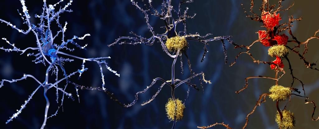
Scientists have revealed that immune cells in Alzheimer’s brains behave differently from those in brains of people without the disease – a discovery that could lead to new treatments.
Published in 2023, an analysis of human brain tissue discovered microglia in the brains of people with Alzheimer’s were more frequently in a pre-inflammatory state, making them less likely to be protective.
Microglia are immune cells that help keep our brains healthy by clearing waste and preserving normal brain function.
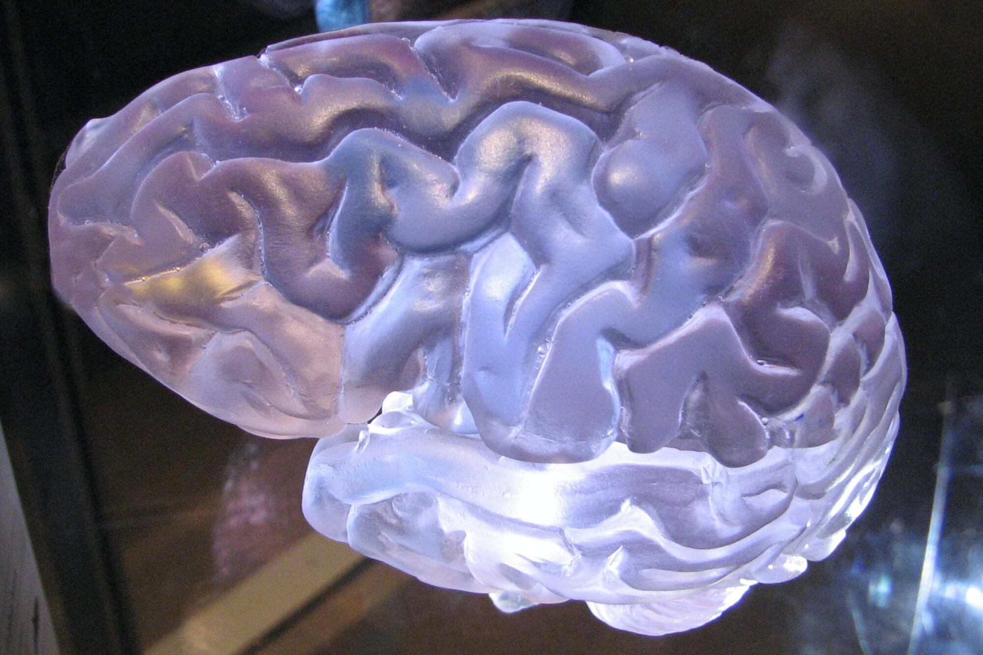
Blocking brain damage triggered by a glioblastoma, an aggressive brain cancer, may slow the growth of the cancer and allow the brain to keep working better for longer, according to a new study led by UCL (University College London) researchers.
The study, published in Nature, looked at glioblastomas in mice. It found that early-stage tumors damaged parts of nerve cells called axons, and that the brain’s natural response to this injury—breaking down and clearing away these damaged axons—accelerated the tumor’s growth.
Mice in whom this natural response was turned off developed less aggressive tumors, lived for longer and maintained normal brain function that persisted to nearly the end of their lives. In contrast, mice who responded to nerve damage as normal developed more aggressive tumors and progressive disability, the researchers found.
Intensity, polarization, and spectrum of light, as distinct dimensional characteristics, provide a comprehensive understanding of light-matter interaction and are crucial across nearly all domains of optical science and technology1,2,3,4. For instance, the polarization information5 is critical for determining material composition and surface texture, whereas spectral analysis is instrumental in medical diagnosis and wavelength-division optical communication6. As modern technology rapidly advances, the demand for comprehensive detection of high-dimensional light field continues to grow7,8.
Conventional detection devices typically measure either spectrum or polarization of input light, sacrificing the valuable information from other dimensions. A common solution is to incorporate multiple discrete diffraction elements and optical filters to separately distinguish light with different polarization and wavelength9,10,11,12. However, this leads to bulky and time-consuming systems. Recently, several integrated high-dimensional detectors based on optical metasurfaces13 have been proposed, and the typical representative relies on mapping different dimensional information into distinct locations, using position and intensity distributions for light field detection14,15. However, as the number of detection parameters increases, the signal crosstalk between different information at different spatial locations become pronounced16,17,18. Another type of detector, based on computational reconstruction, maps light field into a series of outputs, encoding the entire high-dimensional information rather than isolating individual dimension19,20,21,22. Nevertheless, these systems are generally restricted to detecting light fields at a few values with low resolution in each dimension, such as limited polarization and wavelength channels, due to the limited internal degrees freedom in the encoding devices23,24. Additionally, most of them rely on commercial cameras, inevitably requiring numerous detector arrays25. Consequently, achieving fully high-dimensional characterization of arbitrary complex light field with a compact and efficient system remains challenging.
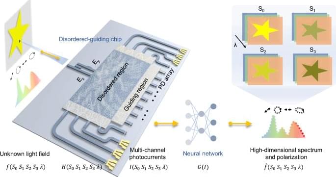
In this work, we propose and demonstrate an on-chip high-dimensional detection system capable of characterizing broadband spectrum along with arbitrary varying full-Stokes polarization through single-shot measurement. The high-dimensional input is encoded into multi-channel intensities through the uniquely designed disordered-guiding chip, and decoded by a multilayer perceptron (MLP) neural network (Fig. 1). The core disordered region introduces complex interference between two separate orthogonal polarization components and multiple scattering to enhance the dispersion effect, enabling rich polarization and spectrum responses. Whereas, the surrounding guiding region based on inverse-design directs the input light to the on-chip photodetectors (PDs), improving the transmittance and detection efficiency. With the assistance of neural network for decoding, we achieve reconstruction of full-Stokes polarization and broadband spectrum with a single measurement. It reveals a high spectral sensitivity of 400 pm with average spectral error of 0.083, and polarization error of 1.2°. Furthermore, we demonstrate a high-dimensional imaging system, exhibiting superior imaging and recognition capabilities compared to conventional single-dimensional detectors. This demonstration holds promising potential for future imaging and sensing applications.