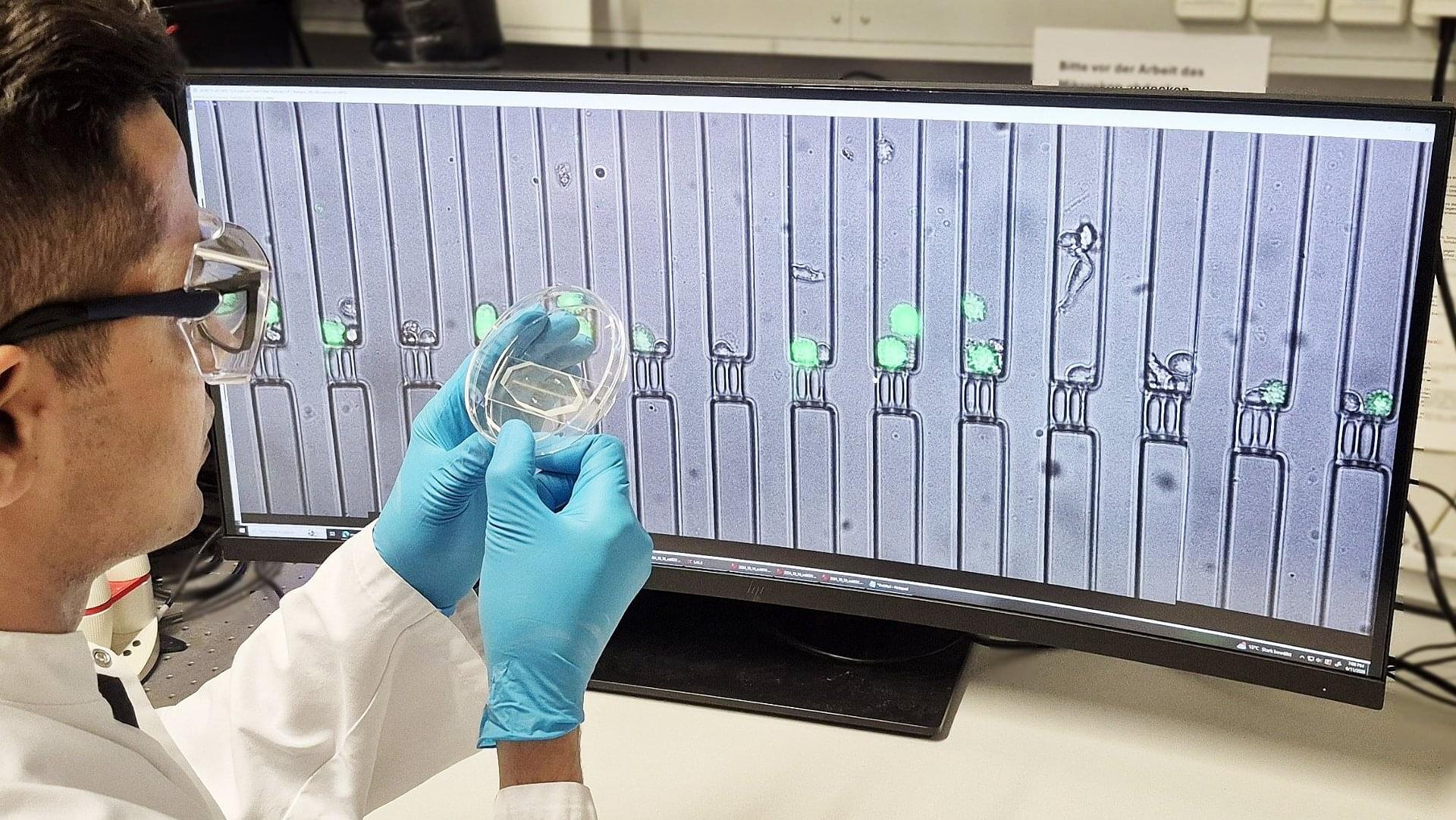
Leipzig University just got the world’s largest brain-inspired supercomputer. Could this be the future of drug discovery?

Li, D., Li, J., Johann, D.J. et al. Augmenting precision medicine via targeted RNA-Seq detection of expressed mutations. npj Precis. Onc. 9, 182 (2025). https://doi.org/10.1038/s41698-025-00993-8
New research by scientists from the U.S. Department of Energy’s Lawrence Berkeley National Laboratory (Berkeley Lab), in collaboration with scientists from TAU Systems Inc., has brought the promise of smaller and more affordable X-ray free-electron lasers one step closer to reality.
X-ray free-electron lasers (XFELs) are powerful light sources and are typically large research instruments. Scientists use them to probe nature’s secrets at the atomic level, enabling advances in medicine, biology, physics, materials, and more. The push to develop more compact and less expensive XFELs is expected to increase the number of facilities that will be able to implement this technology, greatly expanding its impact across many areas of science.
“As part of this effort, we are applying our long-standing expertise in a type of advanced accelerator called laser plasma acceleration to shrink XFELs,” said Sam Barber, a staff scientist in Berkeley Lab’s Accelerator Technology & Applied Physics (ATAP) Division. “In addition to standalone light sources, exceptionally high-quality electron beams from plasma accelerators could be injected into existing XFELs to significantly extend their performance.”
A multi-institutional team led by the Chinese Academy of Sciences has constructed a proteomic atlas of human aging across 13 organs, revealing tissue-specific aging clocks, transcriptome-proteome decoupling, and secreted proteins that may accelerate systemic decline.
Organ-specific aging and deterioration drive vulnerability to chronic diseases. Previous studies focused primarily on plasma proteins or DNA methylation profiles. No investigation has systematically mapped how protein quality control deteriorates differently across tissues identified organ‑specific biological age biomarkers.
In the study, “Comprehensive human proteome profiles across a 50-year lifespan reveal aging trajectories and signatures,” published in Cell, researchers designed a multi-tissue proteomic analysis, charting organ-level protein dynamics and aging-related biomarkers across five decades of adult life to construct a longitudinal proteomic atlas of human aging.
Optimal lesioning is essential for successful MR-guided focused ultrasound (MRgFUS) thalamotomies targeting the ventral intermediate nucleus (Vim) for tremors. This study aimed to evaluate the relationships between postoperative lesions that overlapped with the Vim and surrounding structures segmented automatically and the treatment outcomes.
This study included 48 patients who underwent MRgFUS thalamotomy targeting the Vim for essential tremors. The Clinical Rating Scale for Tremor (CRST) score was examined preoperatively as well as 1 week, 3 months, and 12 months postoperatively. Adverse effects were also assessed 1 month postoperatively. Using automatic segmentation software and fiber tracking software, the authors retrospectively segmented the Vim and surrounding structures, including the internal capsule (IC), ventrocaudal nucleus (Vc), zona incerta (ZI), and dentato-rubro-thalamic tract (DRTT), using preoperative images. Additionally, they manually delineated the coagulated lesions using images taken immediately after MRgFUS thalamotomy. The relationships between the volume and location of lesions overlapping with these structures, CRST improvement rates, and the presence of adverse effects were examined.
The mean thalamotomy volume was 0.076 ± 0.042 cm3 (median 0.085 cm3). The median improvement in the CRST score in the affected upper limb at 12 months postoperatively was 68.8%. Although no correlation was observed between lesion volume and CRST improvement at 1 week postoperatively, a positive correlation was observed between lesion volume and CRST improvement at 3 and 12 months. At 12 months, the authors observed a moderate correlation between the volume of the lesions overlapping with the Vim and improvement in the CRST score. A slightly stronger correlation was observed between the percentage of the lesion volume and the Vim. No correlation was found between lesion volume and improvements in the IC, Vc, ZI, DRTT, or CRST score. However, the authors found that both total lesion volume and the volume of lesion within the IC were significantly associated with gait imbalance.
If it has seemed like more people you know are developing diabetes, you are right. The diabetes epidemic is not called an epidemic for nothing: According to the American Diabetes Association, over 10% of the U.S. population—approximately 38.4 million people—had diabetes in 2021, and 1.2 million more people get diagnosed each year.
Type 2 diabetes occurs when your body develops a resistance to insulin, the hormone that helps regulate glucose levels in your blood. Insulin is secreted by pancreatic cells called β-cells, and in T2D, they ramp up insulin production to try to regulate blood glucose levels, but even that is insufficient and the β-cells eventually become exhausted over time. Thanks to their importance, the functional β-cell mass, or the total number of β-cells and their function, determines a person’s risk of diabetes.
Β-cells are not homogeneous, even within a single individual, and consist of different “subtypes,” each with their own secretory function, viability, and ability to divide. In other words, each β-cell subtype has a different level of fitness, and the higher, the better. When diabetes develops, the proportions of some β-cell subtypes are changed. But a key question remains: Are the proportion and fitness of different β-cell subtypes altered by diabetes or are the changes responsible for the disease?
Is Chief Executive Officer and Chairman of the Board of BioStem Technologies (https://biostemtechnologies.com/), a leading innovator focused on harnessing the natural properties of perinatal tissue in the development, manufacture, and commercialization of allografts for regenerative therapies.
Jason brings a wealth of experience in strategic operations planning and technical projects management from his rigorous technical background. His diverse expertise includes continuous process improvement, training and development programs, regulatory compliance and best practices implementation, and advanced problem solving.
Jason began his career as a technical engineer working for Adecco at SC Johnson in 2009, where he developed comprehensive maintenance plans to support manufacturing processes at scale. He then transitioned to manufacturing and quality engineering for major organizations, including ATI Ladish Forging, Nemak, and HUSCO International, where he spearheaded process design and implementation, solved complex supply-chain and manufacturing problems, and improved product sourcing and purchasing.
Jason’s philanthropic work with the Juvenile Diabetes Research Foundation sparked an interest in biotech, leading him to co-found Biostem Technologies in 2014. As CEO he has leveraged his expertise to optimize tissue sourcing, strategically build out a 6,000 square foot tissue processing facility that is fully compliant with FDA 210,211, 1,271, and AATB standards, and put together an expert team of professionals to support the company’s continued growth.
Jason holds a B.S. in Mechanical Engineering Technology and a minor in Mathematics from the Milwaukee School of Engineering and is Six Sigma Black Belt certified. He also serves as a Processing and Distribution Council Member for the American Association of Tissue Banks (AATB), as well as serves as a member of the Government Affairs committee for BioFlorida.
#JasonMatuszewski #BioStemTechnologies #PerinatalTissue #RegenerativeTherapies #ChronicWounds #DiabeticFootUlcers #VenousUlcers #PressureUlcers #AmnioticTissue #TissueAllografts #ExtracellularMatrix #ECM #GrowthFactors #Cytokines #Collagen #ProgressPotentialAndPossibilities #IraPastor #Podcast #Podcaster #ViralPodcast #STEM #Innovation #Technology #Science #Research
Weill Cornell Medicine researchers have discovered that PD-1—a molecule best known for putting the brakes on immune cells—also plays a critical role in helping T cells become long-term immune defenders in the skin. Early during infection, PD-1 acts like a steering wheel, guiding T cells to become protective resident memory T cells (TRM) that stay in place. These cells remember invading germs or cancer and quickly mount a response if that enemy reappears.
The preclinical findings, published July 29 in Nature Immunology, may impact how clinicians approach cancer treatments called immune checkpoint inhibitors. These drugs bind to PD-1 on the surface of T cells, releasing the brakes and unleashing the immune system to attack cancer cells.
Though immune checkpoint inhibitors are successful in treating melanoma, an aggressive skin cancer, about 40% of patients develop inflammatory rashes and itching in the skin or reactions in other epithelial tissues that cover internal and external surfaces of the body.