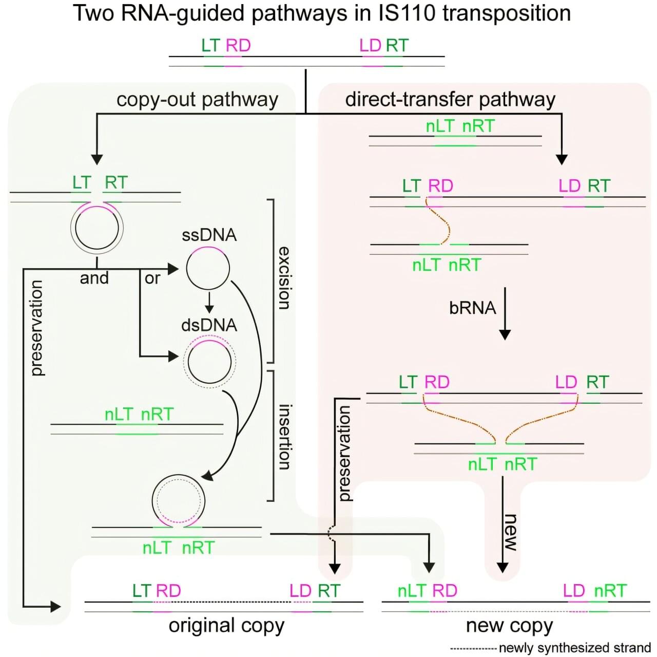
Toggle light / dark theme

‘Three-hit model’ involving genes and environment describes possible causes of autism

A new University of California San Diego School of Medicine study offers a unified biological model to explain how genetic predispositions and environmental exposures converge to cause autism spectrum disorder (ASD).

The study, published in Mitochondrion, describes a “three-hit” metabolic signaling model that reframes autism as a treatable disorder of cellular communication and energy metabolism. The model also suggests that as many as half of all autism cases might be prevented or reduced with prenatal and early-life interventions.

“Our findings suggest that autism is not the inevitable result of any one gene or exposure, but the outcome of a series of biological interactions, many of which can be modified,” said study author Robert K. Naviaux, M.D., Ph.D., professor of medicine, pediatrics and pathology at UC San Diego School of Medicine.

Genetic overlap of 14 psychiatric disorders explains why patients often have multiple diagnoses

An international collective of researchers is delivering new insights into why having multiple psychiatric disorders is the norm rather than the exception. In a study published today in the journal Nature, the team provides the largest and most detailed analysis to date on the genetic roots shared among 14 conditions.

The study is the latest effort from the Psychiatric Genomics Consortium’s Cross-Disorder Working Group, co-chaired by Kenneth Kendler, M.D., a professor in the Department of Psychiatry at Virginia Commonwealth University’s School of Medicine, and Jordan Smoller, M.D., a professor in the Department of Psychiatry at Harvard Medical School.

The majority of people diagnosed with a psychiatric disorder will ultimately be diagnosed with a second or third disorder in their lifetime, creating challenges for defining and treating these conditions. While a person’s environment and lived experience influence their risk for developing multiple disorders, their genetic makeup can also play a significant role.

Stress hormones can alter brain networks and strengthen emotional memories

Stress influences what we learn and remember. The hormone cortisol, which is released during stressful situations, can make emotional memories in particular stronger. But how exactly does cortisol help the brain build emotional memories?

In a new study, Yale researchers investigated just that. Specifically, they wanted to know how cortisol acts separately on brain circuits that track emotion and those that track memory. They found that cortisol not only helped people remember emotional experiences but also enhanced emotional memory by changing the dynamic brain networks associated with both memory and emotion.

“We all experience stress, and my lab is interested in understanding how stress can be helpful,” said corresponding author Elizabeth Goldfarb, an assistant professor of psychiatry at Yale School of Medicine and of psychology in the Faculty of Arts and Sciences.

Polarized light boosts accuracy of wearable health sensors for all skin tones

Photoplethysmography (PPG) is an optical sensing technique that measures blood volume changes and underpins devices ranging from hospital-grade pulse oximeters to consumer wearables that track heart rate, sleep, and oxygenation.

Despite its widespread use, PPG accuracy can vary significantly across individuals, particularly by skin tone. Darker skin contains more melanin, which absorbs and scatters light, often leading to less reliable readings. This disparity has been linked to inaccuracies in blood-oxygen measurements among people with more melanin.

Emerging structural insights into PRC2 function in development and disease

Structural insights into PRC2 function in development and disease.

Polycomb repressive complex 2 (PRC2) is a central epigenetic regulator of developmental gene repression that displays remarkable complexity arising from multiple molecular layers.

Enzyme catalysis and chromatin targeting form the basis of the common and distinct functions of PRC2.1 and PRC2.2, serving as focal points in the cellular regulation of PRC2 activity under both physiological and pathological contexts.

Structural biology has begun to clarify the molecular mechanisms underlying key functions of PRC2 and uncover new modes of regulation, with much still remaining to be understood about the elaborate system of PRC2-mediated gene control. https://sciencemission.com/PRC2-function-in-development-and-disease


Polycomb repressive complex 2 (PRC2) is a key epigenetic enzyme complex that mediates developmental gene repression mainly by depositing the repressive H3K27me3 histone mark. PRC2 operates through its distinct forms, PRC2.1 and PRC2.2, each defined by unique accessory subunits, with additional complexity introduced by other molecular variants such as developmentally regulated homologs and isoforms. PRC2 function is primarily dictated by its enzymatic activity and chromatin recruitment, both of which are rigorously controlled during development and can be dysregulated by disease-associated mutations and oncoproteins. Structural biology has begun to provide important mechanistic insights into various aspects of PRC2 assembly, catalysis, chromatin targeting, and cellular regulation at atomic resolution, addressing several longstanding questions about the Polycomb repression system.

Detrimental Effect of Plasma From Patients With Severe Aortic Stenosis on Valvular Endothelial Cells: Role of Proinflammatory Cytokines and Factor Xa

Severe AS plasma is pro-inflammatory and pro-thrombotic: it drives oxidative stress + endothelial dysfunction + monocyte/platelet adhesion in VECs. Multiple drug classes blunted these effects. @A_Trimaille @adrien_carmona @BMarchandot


Aortic stenosis (AS) is the most prevalent valvular heart disease in developed countries.1 Despite its widespread occurrence, no medical treatment has been validated to prevent the thickening of the aortic valve and the reduction in its opening area. The only available therapeutic option to date remains surgical or transcatheter aortic valve replacement once the severity criteria are met.2, 3 A comprehensive understanding of AS pathophysiology is therefore essential for identifying new therapeutic targets.

Initially thought to be a passive degenerative process, AS is now recognized as an active, multifaceted condition involving numerous cellular and molecular contributors.1, 4 The progression of AS follows a chronological sequence, beginning with the damage and dysfunction of valvular endothelial cells (VECs) due to biomechanical forces acting on the aortic valve. This damage promotes intravalvular inflammation and neoangiogenesis, followed by myofibroblastic and osteoblastic differentiation of valvular interstitial cells. Additionally, several lines of evidence suggest a bidirectional interaction between the pathomechanisms of AS and various components of the hemostatic system, including platelets, tissue factor, thrombin, von Willebrand factor, and extracellular vesicles.4, 5, 6

Given the pivotal role of VECs in maintaining valvular homeostasis under physiological conditions, and their early involvement in AS pathogenesis, they are considered key actors in the disease process. Although biomechanical factors have been identified as primary triggers of VEC dysfunction in the early stage of AS,4 the molecular mechanisms underlying the interaction between plasma from patients with AS and aortic VECs remain unclear. Because plasma contains various biological effectors potentially contributing to endothelial cell dysfunction, including proinflammatory cytokines and hemostatic factors,4, 7 this study aimed to investigate whether plasma from patients with AS induces oxidative stress and contributes to VEC dysfunction.

HSV1716 Prevents Myeloma Cell Regrowth When Combined with Bortezomib In Vitro and Significantly Reduces Systemic Tumor Growth in Mouse Models

Multiple myeloma remains largely incurable due to refractory disease; therefore, novel treatment strategies that are safe and well-tolerated are required.

A Red Blood Cell Protein Turns Dendritic Cells Tolerant

The immune system uses regulatory T cells to dampen inflammatory responses. However, the mechanism that led to these tolerant cells was unclear.

Now, a team of researchers showed that the trigger lies in a protein used in red blood cell production.

Read more.

Erythropoietin, the protein that drives red blood cell formation, also induces tolerance in dendritic cells, leading to the development of regulatory T cells.

Abstract: For those interested in fertility regulation, endometriosis, adenomyosis, and endometrial cancer…

Francesco J. DeMayo & team discover uterine ZMIZ1 co-regulates estrogen receptor to establish and maintain pregnancy and general uterine health via cell growth responses and preventing uterine fibrosis:

The figure shows epithelial cell DNA synthesis (reflected by EdU incorporation) was inhibited by Zmiz1 deletion.


1Pregnancy & Female Reproduction Group, Reproductive and Development al Biology Lab, NIEHS, Research Triangle Park, North Carolina, USA.

2Inotiv-RTP, Durham, North Carolina, USA.

3Division of Hematology-Oncology, Department of Internal Medicine, University of Michigan School of Medicine, Ann Arbor, Michigan, USA.

/* */