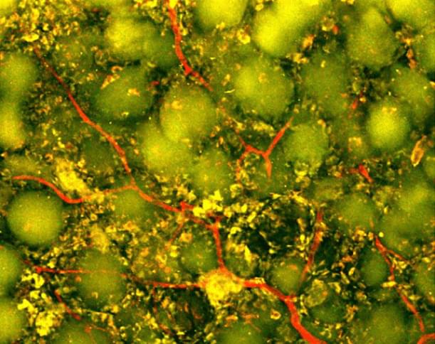
Biomedical engineers at Duke University have developed an injectable biomaterial that helps transform the cavity left behind after a stroke caused by a blood clot into an environment more supportive of repair. By recruiting the body’s own immune cells, the treatment promoted the growth of new blood vessels, supported neural remodeling and improved motor performance in mice. The work appears in Cell Biomaterials.
Every year, millions of people experience strokes caused by blood clots, called ischemic strokes. Immediate treatments, including clot-dissolving drugs and mechanical removal of the clot, can restore blood flow and preserve threatened brain tissue. But these treatments cannot replace tissue that has already been lost.
Large strokes can result in substantial tissue loss and leave behind a cavity at the site of the injury. After the clot is removed, recovery relies largely on rehabilitation, which helps surviving brain circuits adapt but does not directly repair the damaged area.
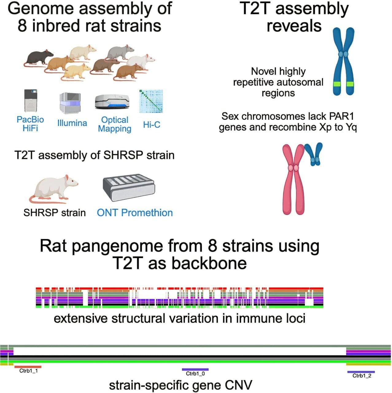
Researchers have created the most complete genetic profile of the brown rat to date, according to a UTHealth Houston-led team, paving the way for scientists to more accurately investigate genetic links to conditions like heart disease, kidney disease, high blood pressure and stroke.
The research, published in Cell Genomics, was led by corresponding author Peter Doris, Ph.D., director of the Center for Human Genetics at The Brown Foundation Institute of Molecular Medicine within McGovern Medical School at UTHealth Houston.
The assembly of the brown rat’s genome provides a complete genetic fingerprint and reveals that the brown rat’s DNA is more complex than scientists previously understood. In addition to uncovering more than 60 new genes, many of which were previously difficult to sequence and are thought to play a role in immunity and other biological processes, the team discovered that brown rat sex chromosomes differ significantly from those in humans.
For most of human history, scientists believed that once a cell became a skin cell, a neuron, or a heart cell, that identity was permanent. Then a group of researchers discovered something extraordinary: cells could be reset. My guest today was there when that discovery happened.
Dr. Koji Tanabe, Ph.D. is Founder and CEO of I Peace (https://ipeace.com/en/), one of the world’s leading companies advancing induced pluripotent stem cell — or iPSC — technology from the research laboratory into scalable clinical manufacturing.
Dr. Tanabe occupies a truly unique place in modern biomedical history. He earned his Ph.D. in the laboratory of Nobel Laureate Dr. Shinya Yamanaka at Kyoto University and was the second author on the landmark scientific paper that first demonstrated the successful creation of human induced pluripotent stem cells — a discovery that fundamentally changed regenerative medicine and ultimately earned Dr. Yamanaka the 2012 Nobel Prize.
After helping establish one of the most important technologies in modern biology, Dr. Tanabe continued his work at Stanford University in the laboratory of Dr. Marius Wernig, a pioneer of direct cellular reprogramming, where he investigated how mature blood cells can be directly converted into neurons and explored the molecular mechanisms that govern cellular identity.
In 2015, Dr. Tanabe founded I Peace with an ambitious vision: to make clinical-grade iPS cells accessible at industrial scale. Today, the company has developed automated GMP manufacturing platforms capable of producing personalized and clinical-grade iPS cells for researchers, pharmaceutical companies, and regenerative medicine programs around the world.
On the episode we’ll explore how far the field has come since those first groundbreaking experiments nearly two decades ago, where regenerative medicine stands today, the growing role of iPS cells in drug discovery and transplantation, the excitement surrounding in vivo reprogramming and partial cellular rejuvenation, and what may ultimately become possible when every individual has access to their own personalized stem cell bank.
Basically whole body regeneration is definitely possible we just need to right genetic code to push the regeneration button in the human body much like how these mice had their digits regenerated so too we can regenerate just like Deadpool or even the axolotl.
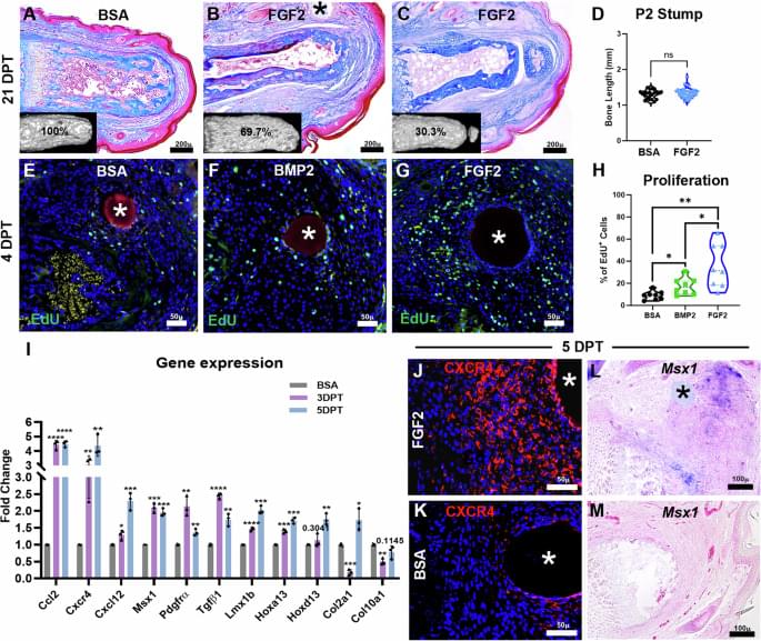
Wound fibrosis after amputation in mammals is replaced with regeneration of amputated structural elements by sequential FGF2/BMP2 treatment. Regenerated tissues include phalangeal/sesamoid bones, tendon/ligament, synovial joint, articular cartilage.
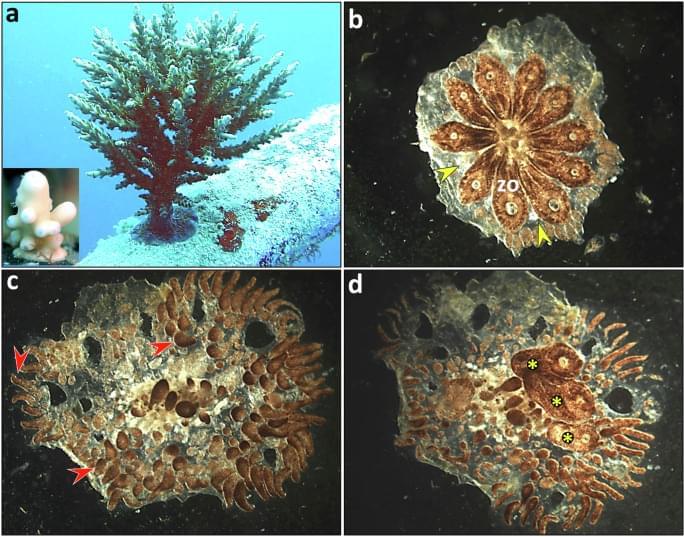
Whole body regeneration is possible just would need to find it in human beings similar genetics.
Rinkevich, Y., Rinkevich, B. From fragment to form: whole-body regeneration in a model urochordate. npj Regen Med10, 36 (2025). https://doi.org/10.1038/s41536-025-00423-0
Background and ObjectivesMigraine is a highly prevalent and disabling neurologic disorder. Beyond drug-specific mechanisms, the role of contextual, individual-related, and disease-related factors remains poorly characterized. We quantified placebo…
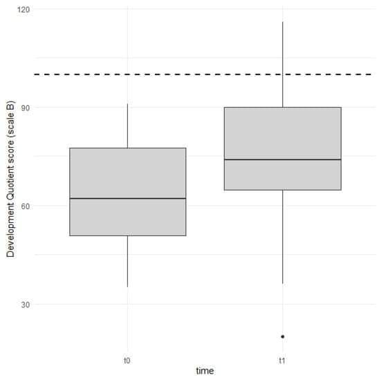
Background/Objectives: The primary objective of the present study was to investigate early global development in children after one year of cochlear implant (CI) use. The secondary objective was to investigate the role of variables such as age at CI activation, gender, and parental schooling in early global development in children with a CI. Methods: The study sample included 24 subjects. All children were affected by severe-to-profound congenital bilateral sensorineural hearing loss (HL). The HL was diagnosed between 1 and 23 months of age (median 3 months) and participants underwent cochlear implant activation at 9–25 months (median 14 months). Participants were evaluated before CI surgery and after one year of CI use using the Italian version of the Griffiths III scales.
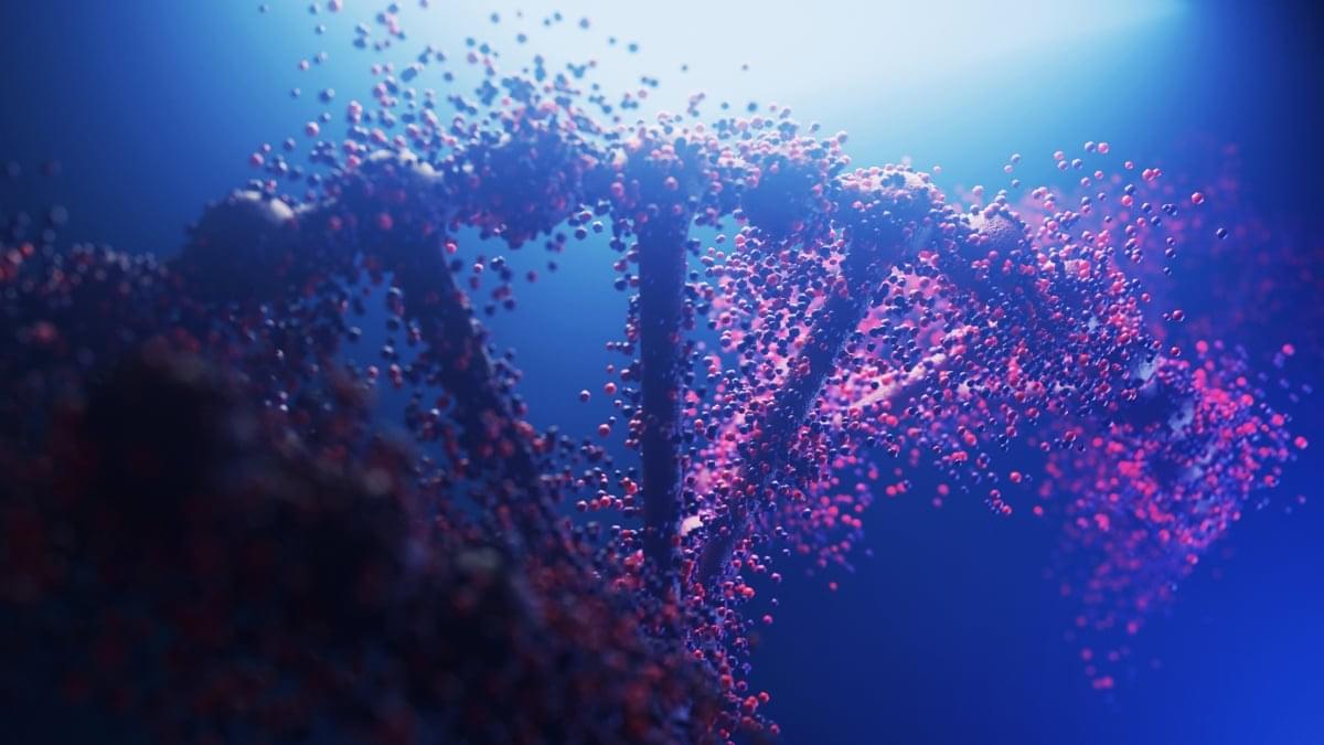
One of the most iconic visual representations in biological science is the double helix – the twisting, twin-stranded ladder that defines the shape of DNA molecules.
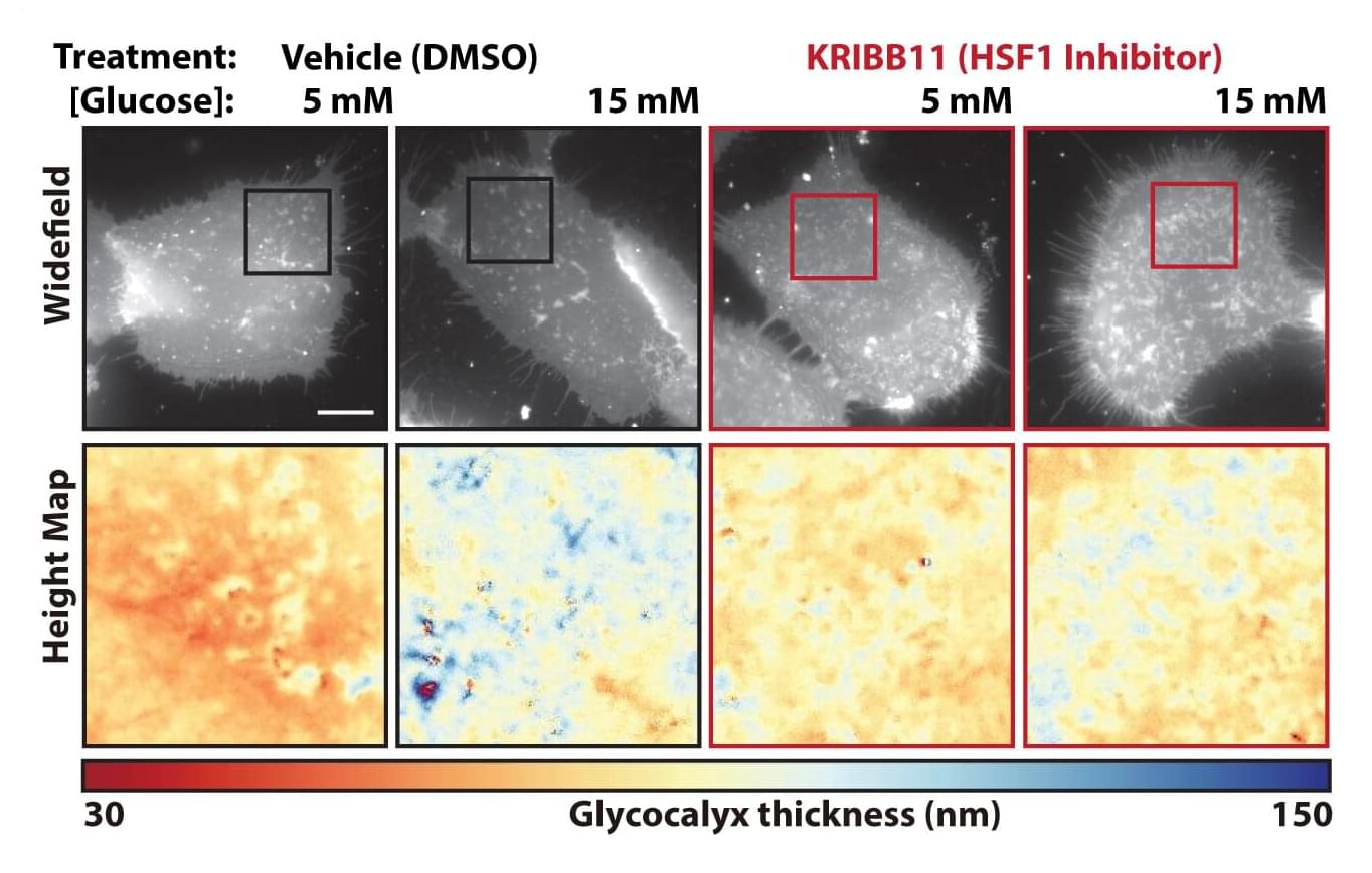
Like spies evading detection by mastering disguises, many cancer cells are adorned with a copious coat of sugar-derived molecules that throws the proverbial hounds of the immune system off the scent.
Scientists at Sanford Burnham Prebys Medical Discovery Institute and collaborators across North America published findings Aug. 7, 2026, in Science Advances showing that cancer cells’ cloaking costumes can result from changes in the nearby neighborhood of immune cells, connective tissue, blood vessels, proteins and carbohydrates called the tumor microenvironment. The researchers also found a way to thin this sugary shroud, enabling cancer cells to be recognized and eliminated by the immune system.
Lead and corresponding author Kevin Tharp, Ph.D., knew that cells squeezed by their surroundings change their mitochondrial function in surprising ways. He realized that a key place where cells would experience this kind of physical pressure was in the tumor microenvironment.
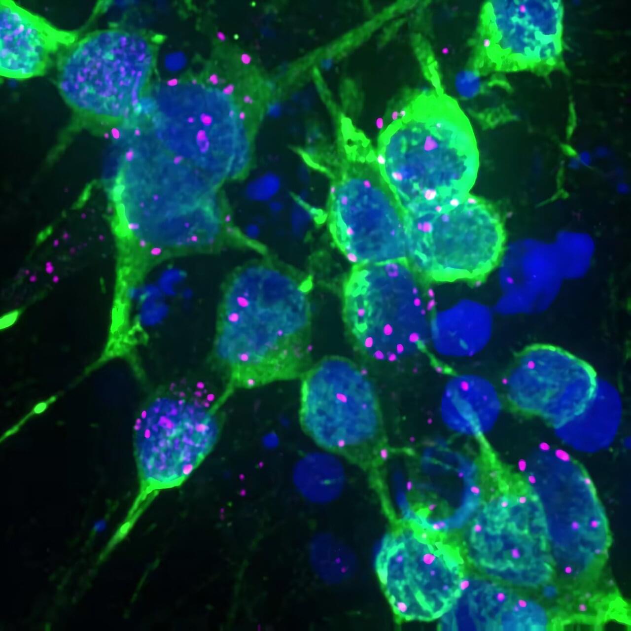
Parkinson’s disease affects more than 10 million people worldwide. The disease is characterized by the buildup of abnormal clumps of the protein alpha-synuclein inside brain cells, but scientists have long struggled to understand exactly how these toxic forms of the protein cause neurons to malfunction and eventually die.
Researchers from the Tofaris lab, part of the Nuffield Department of Clinical Neurosciences and based in the Kavli Institute for Nanoscience Discovery, combined advanced molecular analyses of human stem cell models of Parkinson’s disease with studies of postmortem brain tissue from people with Parkinson’s disease to investigate the earliest stages of the disease process.
The study is published in the journal Nature Communications.