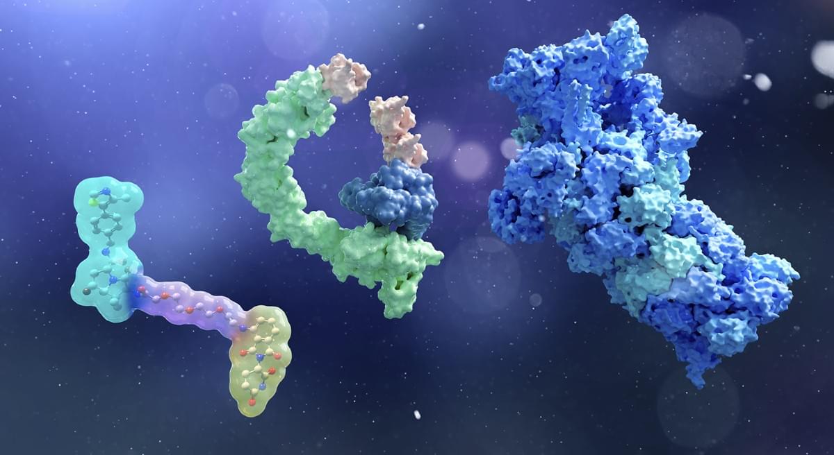
An innovative funding initiative in South Korea has the ambitious goal of expanding the range of potential ‘druggable’ protein targets by pursuing unprecedented therapeutic approaches.

Because glucosamine is widely available and frequently used by older adults to support joint health, the researchers wanted to determine whether it could influence Alzheimer’s disease and related dementias (ADRD).
Working with collaborators Yi Guo, Ph.D., and Jiang Bian, Ph.D., the team used artificial intelligence to analyze deidentified UF Health records collected between 2012 and 2024. They focused on patients diagnosed with either ADRD or mild cognitive impairment (MCI).
Among those patients, researchers found that glucosamine use was relatively common. A total of 1,896 patients with ADRD and 2,750 patients with MCI reported taking the supplement, representing about 8% of each group.
Two of my favorite people. Definitely worth a view if you are interested in either.
Few thinkers have shaped our understanding of the future as profoundly as Ray Kurzweil. An American inventor, computer scientist, futurist, entrepreneur, and bestselling author, Kurzweil is widely regarded as one of the most influential technological forecasters of our time. For decades, he has accurately predicted many of the innovations that now define modern life, from mobile computing and artificial intelligence to digital assistants and large language models often years before they entered the mainstream.
In this special conversation, Tony Robbins sits down with Ray Kurzweil in San Francisco to explore one of the most important questions facing humanity: What happens next?
Together, they examine the accelerating pace of artificial intelligence, the path toward Artificial General Intelligence (AGI), the rise of autonomous agents, the future of work and education, breakthroughs in healthcare and longevity, and how these technologies may transform society over the coming decade.
Kurzweil explains why his long-standing prediction of AGI by 2029 now appears increasingly conservative, why the next few years may bring more change than any period in human history, and how humanity may ultimately merge with the very technologies it creates.

Boston-based biotechnology company Life Biosciences has launched the first-in-human clinical trials of a pioneering “partial cellular reprogramming” technique designed to treat optic nerve damage caused by glaucoma and NAION. Based on previous genetic research, the therapy utilizes a modified virus to deliver three youth-restoring genes to retinal cells, aiming to reverse cellular aging while preserving their specialized functions. Addressing the critical risk of inducing cancer through uncontrolled cell division, the protocol incorporates a vital safety switch: the rejuvenating genes are only activated in the presence of the antibiotic doxycycline. The eye was strategically selected for these initial trials because its relative isolation minimizes the risk of systemic, life-threatening side effects. Involving up to 12 patients, this groundbreaking study serves as a crucial test not only for the potential restoration of vision but for the safety, viability, and future reputation of partial reprogramming as a broader anti-aging and regenerative medicine therapy.
A participant in a landmark clinical trial has been given a cellular-reprogramming treatment that aims to rejuvenate damaged cells in the eye.

Every day, across thousands of American hospitals, artificial intelligence quietly shapes decisions that determine patient outcomes. An algorithm flags a patient as high risk for sepsis; a risk score informs whether a woman receives additional cancer screening; a deterioration model triggers an alert that sends a care team to a bedside. These tools are embedded in the workflows of nearly two-thirds of US hospitals, integrated into the electronic health record systems clinicians rely on daily. But many have never been reviewed by the FDA.
A new viewpoint in The Lancet Digital Health, co-authored by researchers at MIT’s Computer Science and Artificial Intelligence Laboratory (CSAIL) and Jameel Clinic, traces how this problem took root, why it carries serious consequences, and what genuine transparency would require to fix it.
The argument, the scientists say, is not that AI has no place in clinical decision-making. It is that a $4 billion market of clinical decision support tools operates largely beyond public accountability, leaving patients and providers often unable to know whether the tools influencing their care have been validated, by whom, or for which populations they work as intended.
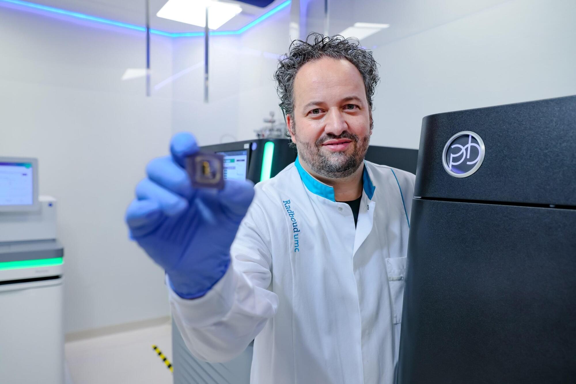
A new test provides a much more complete picture of DNA than current standard diagnostics and leads to a diagnosis more often. The test can replace 15 other tests, making it faster and more efficient. Researchers from Radboud university medical center recommend in the New England Journal of Medicine that this test be adopted everywhere as the first choice for rare genetic disorders.
A condition is considered rare if it affects fewer than 1 in 2000 people. Nevertheless, up to 400 million people worldwide have a rare disease, as there are more than 7,000 different types. Eighty percent of these have a genetic cause. A diagnosis often takes years to obtain. Yet a diagnosis is important: It provides clarity, insight into the future, contact with others in similar situations, and the possibility to assess risks when planning to have children.
Researchers from Radboudumc and Maastricht UMC+ are working together to increase the chances of diagnosing genetic disorders. They compared current standard diagnostics—often involving multiple tests to reach a diagnosis—with a new DNA test in 1000 patients.
To learn more, please visit the YouTube Help Center: https://www.youtube.com/help