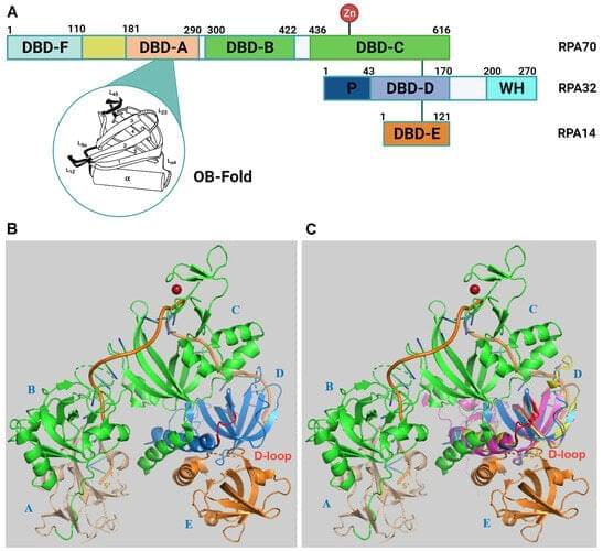
Replication protein A (RPA) is a heterotrimeric protein complex and the main single-stranded DNA (ssDNA)-binding protein in eukaryotes.
When a cell divides, it performs a feat of microscopic choreography—duplicating its DNA and depositing it into two new cells. The spindle is the machinery behind that process: It latches onto chromosomes (where DNA is stored) and separates them so they can settle into their new homes. This tricky process can sometimes go wrong, causing infertility, genetic disorders, or cancer.
Scientists have a good understanding of what spindles are made of: long, thin rods called microtubules as well as a variety of associated motor proteins. However, how these microtubules interact and organize to guide the spindles’ function has remained a mystery.
One approach to understand how the spindle self-organizes is to treat it like an active liquid crystal. Liquid crystals, like spindles, are made up of elongated subunits. Unlike liquid crystals in LCD displays, which require an external electric field to reorient their subunits, spindles are active materials that generate forces internally.
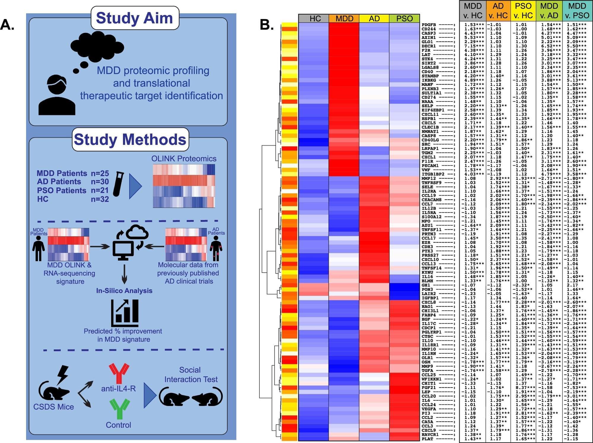
A team of leading clinical research scientists from the Departments of Psychiatry and Dermatology at the Icahn School of Medicine at Mount Sinai has found that the serum of patients with major depressive disorder shares immune abnormalities with inflammatory skin diseases, most notably the common Th2 immune pathway that is implicated in atopic dermatitis. Because these skin diseases are treatable, the findings suggest new therapeutic possibilities for psychiatric illness as well.
The study findings, published in Molecular Psychiatry, underscore the potential role of the Th2 axis in major depressive disorder and highlight the potential of targeting this specific immune pathway that involves interleukin-4 receptor alpha, a cell receptor known to play a key role in regulating inflammation, as a disease-modifying treatment for this psychiatric disorder.
Furthermore, the back-translational drug repurposing strategy employed in this study may offer a new approach to identifying immunomodulatory drugs in psychiatry.
What happens to soft matter when gravity disappears? To answer this, UvA physicists launched a fluid dynamics experiment on a sounding rocket. The suborbital rocket reached an altitude of 267 km before falling back to Earth, providing six minutes of weightlessness.
In these six minutes, the researchers 3D-printed large droplets of a soft material similar to the inks used for bioprinting —a developing technology that shows huge potential for regenerative and personalized medicine, tissue engineering and cosmetics. Bioprinting involves 3D-printing a mix of cells and bio-inks or bio-materials in a desired shape, often to construct living tissues.
The experiment was called COLORS (COmplex fluids in LOw gravity: directly observing Residual Stresses). Using a special optical set-up, the researchers could see where the printed material experienced internal stresses (forces) as the droplets spread and merged. Stressed regions stand out as bright colors in the experiment. Investigating how and where these stresses emerge is important because they can get frozen in a material as it solidifies, creating weak points where 3D-printed objects are most likely to break.
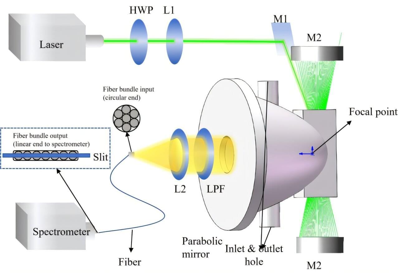
A research team led by Prof. Fang Yonghua from the Hefei Institutes of Physical Science of the Chinese Academy of Sciences proposed and systematically optimized a novel parabolic mirror cavity-enhanced Raman spectroscopy (PMCERS) technique, achieving a marked improvement in gas detection sensitivity through the integration of advanced optical design and signal processing methods. These results were published in Optics & Laser Technology.
Multi-component gas detection is important for environmental, industrial, and medical applications. Raman spectroscopy is well-suited for this purpose because it enables the simultaneous, water-vapor-free detection of multiple gas species. However, its inherently weak scattering limits sensitivity. Conventional cavity-enhanced approaches relying on lens-based collection have a limited numerical aperture, resulting in inefficient capture of three-dimensionally distributed Raman signals.
In this study, the team developed a parabolic mirror-based cavity-enhanced Raman spectroscopy system that leverages the large-aperture characteristics of parabolic mirrors to significantly improve Raman signal collection. Through the systematic optimization of the cavity structure, an efficient closed-loop optical path was established, effectively eliminating signal collection blind spots and suppressing stray-light interference.
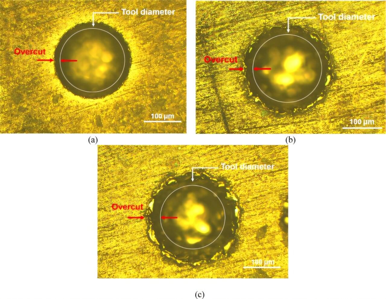
Researchers have developed a new machine-learning-assisted approach to optimize micro-electro-discharge machining (µ-EDM) of a next-generation biocompatible titanium alloy, potentially improving the manufacturing of advanced medical and aerospace components.
The work is published in the journal Scientific Reports.
Titanium alloys are widely used in biomedical implants, aerospace systems, and automotive engineering due to their strength, corrosion resistance, and low weight. However, the commonly used alloy Ti–6Al–4V contains aluminum and vanadium, elements associated with long-term toxicity risks in biomedical applications.
WASHINGTON (AP) — Scientists are testing an entirely new way to fight heart disease: a gene-editing treatment that might offer a one-time fix for high cholesterol.
It’s very early stage research, tried in only a few dozen people so far. But gene-editing approaches being developed by two companies show hints that switching off certain genes could dramatically lower artery-clogging cholesterol, raising hopes of one day being able to prevent heart attacks without having to take pills.
“People want a fix, not a bandage,” said Dr. Luke Laffin, a preventive cardiologist at the Cleveland Clinic. After co-authoring a promising study published in the New England Journal of Medicine, he said he was flooded with queries about how to participate in the next clinical trial.
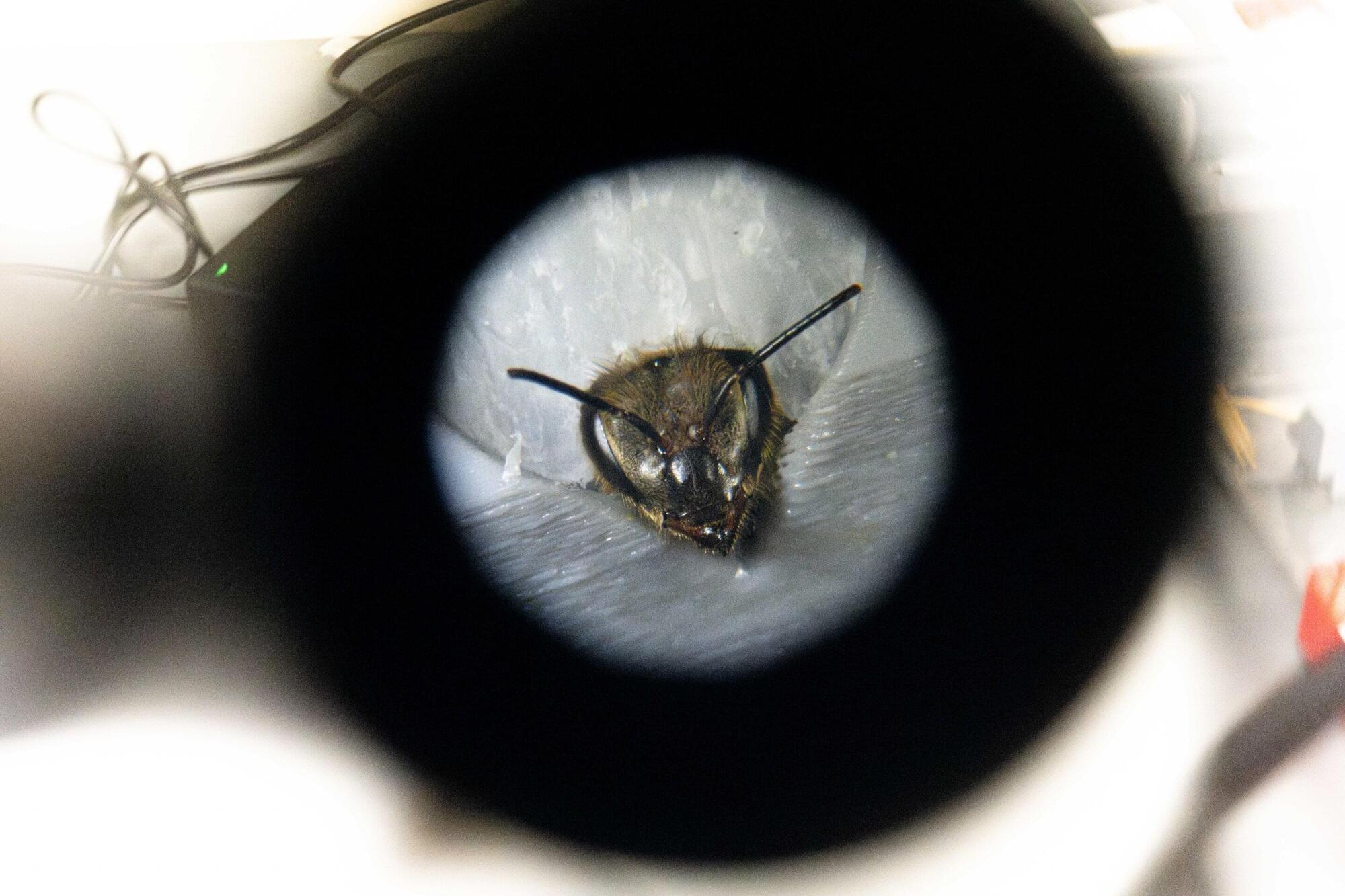
A multi-institutional team of researchers led by Virginia Tech’s Fralin Biomedical Research Institute at VTC has for the first time identified specific patterns of brain chemical activity that predict how quickly individual honey bees learn new associations, offering important insights into the biological basis of learning and decision-making. The study, published in Science Advances, found that the balance between the neurotransmitters octopamine and tyramine can predict whether a bee will learn quickly, slowly, or not at all, as they associate an odor with a reward.
Because the same ancient brain chemicals that guide learning in bees also shape attention and learning in people, the findings may help scientists better understand why individual humans learn at different speeds—and how those processes may go awry in a variety of brain disorders.
Specific patterns of brain chemical activity appear before learning begins and again when a learned behavior first emerges, signaling how quickly an individual bee will learn. The research can help explain how chemicals in the brain drive attention and reinforce learning, with implications for fundamental biology, medicine, and agriculture.

Found in everything from protein bars to energy drinks, erythritol has long been considered a safe alternative to sugar.
But research suggests this widely used sweetener may be quietly undermining one of the body’s most crucial protective barriers – with potentially serious consequences for heart health and stroke risk.
A study from the University of Colorado suggests erythritol may damage cells in the blood-brain barrier, the brain’s security system that keeps out harmful substances while letting in nutrients.