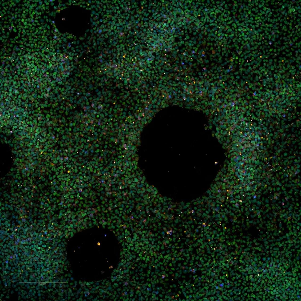
Stem cell–derived islets can be enriched by density gradient separation.

Is Chief Executive Officer and Managing Director of Cynata Therapeutics (https://cynata.com/), a stem cell and regenerative medicine company that is known for its proprietary Cymerus platform, for the scalable and consistent production of mesenchymal stem cell (MSC)-based therapies.
Unlike traditional MSC therapies that rely on multiple donors, the Cymerus manufacturing process ensures that cells for therapeutic use can be produced in virtually limitless quantities from a single donor – making the opportunities endless and attractive from a manufacturing standpoint. The company has completed Phase I studies for Graft vs Host disease & Diabetic Foot Ulcers and have a number of Phase II, and even have a Phase III clinical trial, in progress.
Dr. Kelly has over 20 years’ experience in biopharmaceutical research and development, including almost 15 years focused on the development of mesenchymal stem cell (MSC) based therapies. He joined Cynata in March 2014, initially as Vice President, Product Development, then Chief Operating Officer from May 2019, and since July 2023 has been CEO & MD. At Cynata, he has overseen all stages of the development of the Cymerus induced pluripotent stem cell (iPSC)-derived MSC technology, including the first completed clinical trial of any iPSC-derived product worldwide.
Dr. Kelly previously held positions at Biota Pharmaceuticals, Mesoblast Limited, Kendle International, Amgen and AstraZeneca.
Dr. Kelly holds a Masters in Pharmacy degree from the Robert Gordon University, Aberdeen, a Ph.D. in Pharmaceutical Sciences from Strathclyde University, Glasgow, and he is a Graduate of the Australian Institute of Company Directors (AICD), Melbourne. He is a member of the International Society for Cell and Gene Therapy (ISCT), the International Society for Stem Cell Research (ISSCR), the Royal Pharmaceutical Society and the AICD.
Dr. Kelly also serves on the ISCT Asia-Pacific Industry Committee, the ISSCR Best Practices Working Group for the Development of PSC-Derived Therapies and the Industry Interface Committee of the Center for Commercialization of Regenerative Medicine (CCRM) Australia.
Plastics are everywhere—from packaging and textiles to electronics and medical devices. As plastic waste breaks down, it releases microscopic particles that can penetrate our ecosystems, hinder plant growth, and potentially transfer harmful pollutants to organisms, including humans. Therefore, these plastic particles are a potential threat to the ecosystem, especially in their nanoparticulate form (1–100 nm diameter), which can penetrate the environment through different routes, including the soil beneath our feet.
With this in mind, a team of researchers from Japan set out to study the migration behavior of nanoplastics in different soil types. The study was led by Kyouhei Tsuchida, a Ph.D. student from the National Institute of Advanced Industrial Science and Technology (AIST) and Waseda University, Japan, with fellow students Yukari Imoto, Takeshi Saito, and Junko Hara also from AIST, and Professor Yoshishige Kawabe from the Department of Resources and Environmental Engineering, Waseda University. This study was published online in the journal Science of the Total Environment on April 4, 2025.
The researchers focused on the adsorption of the nanoplastics on soil and the aggregation characteristics of both the nanoplastics and soil particles under varying pH conditions. “The aggregation properties of nanoplastics and their adsorption onto soil particle surfaces are known to affect their migration in soil,” notes Tsuchida. “We conducted experiments to analyze these traits to get a better understanding of the migration of nanoplastics.”
A stem cell treatment helped improve the motor function of two out of four patients with a spinal cord injury in the first clinical study of its kind, Japanese scientists said.
There is currently no effective treatment for paralysis caused by serious spinal cord injuries, which affect more than 150,000 patients in Japan alone, with 5,000 new cases each year.
Researchers at Tokyo’s Keio University are conducting their study using induced pluripotent stem cells (iPS)—created by stimulating mature, already specialized, cells back into a juvenile state.
A Japanese university said Friday it has successfully transplanted stem cells into a patient with a spinal cord injury, in the first clinical trial of its kind.
There is currently no effective treatment for paralysis caused by serious spinal cord injuries, believed to affect more than 100,000 people in Japan alone.
Surgeons at Tokyo’s Keio University want to study whether induced pluripotent stem (iPS) cells can be used to treat the injuries.
Lithium was introduced into psychiatric practice in the late nineteenth century and has since become a standard treatment for severe psychiatric disorders, particularly those characterized by psychotic agitation. It remains the most effective agent for managing acute mania and preventing relapses in bipolar disorder. Despite potential adverse effects, lithium’s use should be carefully considered relative to other treatment options, as these alternatives may present distinct safety and tolerability profiles. The World Health Organization classifies lithium salts as ‘essential’ medications for inclusion in global healthcare systems. Over the past two decades, the growing recognition of lithium’s efficacy—extending beyond mood stabilization to include reducing suicide risk and inducing neuroprotection—has led to its incorporation into clinical practice guidelines.
Key to this innovation in ultrasound imaging—a method called Nonlinear sound sheet microscopy —was the discovery of a sound-reflecting probe. The author said: “This probe is a nanoscale gas-filled vesicle that lights up in ultrasound images, making cells visible. These vesicles have a protein shell and we can engineer them to tune their brightness in images. We used these gas vesicles to track cancer cells.”
In addition to revealing cells, the team used ultrasound and microbubbles as probes circulating in the blood stream to detect brain capillaries. The author said: “To our knowledge, nonlinear sound sheet microscopy is the first technique capable of observing capillaries in living brains. This breakthrough has tremendous potential to diagnose small vessel diseases in patients.” Since microbubble probes are already approved for human use, this technique could be deployed in hospitals in a few years.
Ultrasound is one of the most widely used imaging techniques in medicine, but up until recently it hardly played a role in imaging the tiniest structures of our bodies such as cells. “Clinical ultrasound, like the kind used for pregnancy scans, creates real-time images of body parts”, the first author explains. “It allows diagnosis of various diseases, or to monitor a developing baby. However, what is going on at a microscopic level remains hidden.”
Now, a team of scientists managed to image specifically labelled cells in 3D with ultrasound. For the first time, they imaged living cells inside whole organs across volumes the size of a sugar cube. In comparison, current light-based microscopes often require imaging of non-living samples, the author says. “The sample or organ of interest has to be removed and processed, and you lose the ability to track activity of cells over time”
First, the team discovered that heparan sulfate (HSPG), a sulfated glycoprotein on the cell surface, plays a crucial role in attracting LNPs and facilitating mRNA entry into the cell.
- Second, they identified V-ATPase, a proton pump at the endosome, which acidifies the vesicle and causes LNPs to become positively charged, enabling them to temporarily disrupt the endosomal membrane and release the mRNA into the cytoplasm to be expressed.
- Lastly, the study uncovered the role of TRIM25, a protein involved in the cellular defense mechanism. TRIM25 binds to and induces the rapid degradation of exogenous mRNAs, preventing their function.
So how do the mRNA vaccines evade this cellular defense? A key finding of the study was that mRNA molecules containing a special modification called N1-methylpseudouridine (m1Ψ)—which was awarded the 2023 Nobel Prize in Physiology or Medicine—can evade TRIM25 detection. This modification prevents TRIM25 from binding to mRNA, enhancing the stability and effectiveness of mRNA vaccines. This discovery not only explains how mRNA vaccines evade cellular surveillance mechanisms but also emphasizes the importance of this modification in enhancing the therapeutic potential of mRNA-based treatments.
Additionally, the research highlighted the critical role of proton ions in this process. When the LNPs rupture the endosomal membrane, proton ions are released into the cytoplasm, which activates TRIM25. These proton ions act as a signal that alerts the cell to the invading foreign RNA, which in turn triggers a defense response. This is the first study to demonstrate that proton ions serve as immune signaling molecules, providing new insights into how cells protect themselves from foreign RNA.
A team of researchers has uncovered a key cellular mechanism that affects the function of mRNA vaccines and therapeutics. Their study, recently published in Science, provides the first comprehensive understanding of how mRNA vaccines are delivered, processed, and degraded within cells—a breakthrough that could pave the way for more effective vaccines and RNA-based treatments.
Researchers at Ben-Gurion University of the Negev have developed a machine-learning algorithm that could enhance our understanding of human biology and disease. The new method, Weighted Graph Anomalous Node Detection (WGAND), takes inspiration from social network analysis and is designed to identify proteins with significant roles in various human tissues.
Proteins are essential molecules in our bodies, and they interact with each other in complex networks, known as protein-protein interaction (PPI) networks. Studying these networks helps scientists understand how proteins function and how they contribute to health and disease.
Prof. Esti Yeger-Lotem, Dr. Michael Fire, Dr. Jubran Juman, and Dr. Dima Kagan developed the algorithm to analyze these PPI networks to detect “anomalous” proteins—those that stand out due to their unique pattern of weighted interactions. This implies that the amount of the protein and its protein interactors is greater in that particular network, allowing them to carry out more functions and drive more processes. This also indicates the great importance that these proteins have in a particular network, because the body will not waste energy on their production for no reason.