Toggle light / dark theme

Proton therapy for colorectal cancer

SA-CME credits are available for this article here.

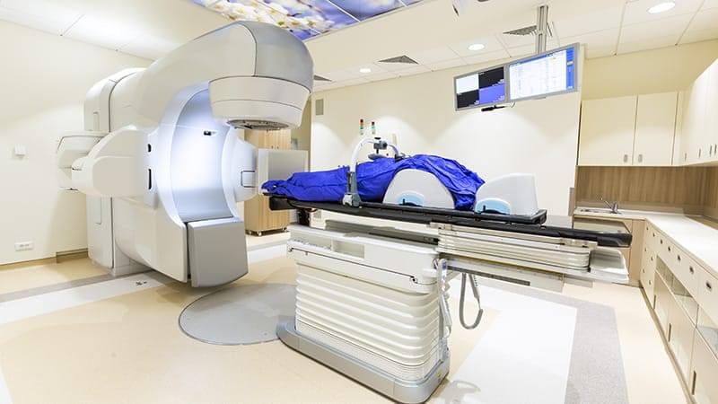
Ever since the first proton beam therapy (PBT) treatment in 1954 at University of California, Berkley, the use of PBT worldwide has rapidly increased.1 Due to the depth-dose characteristics of protons that allow for steep fall-off just distal to the tumor target, PBT can reduce unnecessary radiation dose to nearby normal tissues and allow for safer dose escalation in select clinical scenarios. Superior normal tissue avoidance can lead to reductions in acute and late toxicities, safe dose escalation can lead to improved local control, and the combination of both factors has the potential to impact overall survival (OS).

Early data have suggested that PBT led to improved clinical outcomes in the treatment of various pediatric cancers, ocular melanomas, sarcomas of the paravertebral region, and brain tumors when compared with traditional photon-based radiation.2 Historically, fewer studies evaluated the utility of PBT in the treatment of gastrointestinal (GI) malignancies; however, retrospective studies in the setting of gastroesophageal cancer and pancreatic cancer show that preoperative PBT may reduce postoperative complications and definitive PBT may improve outcomes for those with unresectable disease.3–6 Even fewer studies have evaluated the role of PBT in the primary or neoadjuvant treatment of colorectal cancer (CRC), but there have been published clinical outcomes in the treatment of recurrent disease as well as liver metastases. The aim of this review is to discuss the existing dosimetric and clinical data for PBT in the treatment of patients with CRC.

Injectable ‘satellite livers’ could offer an alternative to liver transplantation

More than 10,000 Americans who suffer from chronic liver disease are on a waitlist for a liver transplant, but there are not enough donated organs for all of those patients. Additionally, many people with liver failure aren’t eligible for a transplant if they are not healthy enough to tolerate the surgery.

To help those patients, MIT engineers have developed “mini livers” that could be injected into the body and take over the functions of the failing liver.

In a new study in mice, the researchers showed that these injected liver cells could remain viable in the body for at least two months, and they were able to generate many of the enzymes and other proteins that the liver produces.

Chemically ‘stapled’ peptides used to target difficult-to-treat cancers

Researchers at the University of Bath have developed a new technology that uses bacteria to build, chemically stabilize, and test millions of potential drug molecules inside living cells, making it much quicker and easier to discover new treatments for difficult-to-treat cancers.

Scientists based at the University’s Department of Life Sciences are investigating peptides—short chains of amino acids, the building blocks of proteins—as potential drugs for a family of notoriously “undruggable” cancer drivers known as transcription factors. These proteins act as master switches that control gene activity and are frequently overactive in cancer.

Swarming, spinning microrobots can manipulate their surroundings

E pluribus unum – “out of many, one” – is not only a motto for the United States. It’s a good credo for microrobots.

A research collaboration between Cornell and the Max Planck Institute for Intelligent Systems has shown how a swarm of microrobots spinning on a water surface can together generate the fluidic torque needed to manipulate passive structures without any physical contact.

This collective behavior was demonstrated to operate gears and move objects, with the aim of eventually performing microscale tasks and biomedical procedures.

Cancer stress protein helps tumors hide from immunity

The ISR in cancer cells triggers the production of a protein called Activating Transcription Factor 4, or ATF4, which in turn triggers the action of many genes that help cancer cells survive, the study authors say. The new work shows that ATF4 also instructs the cell to release LCN2 to protect the tumor from the immune system.

The research team found that LCN2 passes on the ATF4 message to switch macrophages, a type of immune cell abundant in tumors, into an “immunosuppressive” mode, which keeps cancer-killing T cells from entering the tumor.

Whereas ATF4 operates inside cancer cells, LCN2 is released outside where it can be more easily targeted by drugs, the researchers said. Therefore, they designed an antibody therapy, a lab-made version of an immune protein, to bind and block LCN2, which kept it from manipulating macrophages, letting the sidelined T cells back into tumors.

When the researchers team engineered mice to both develop cancer, and to lack LCN2, tumor growth slowed. That this effect happened only in mice with healthy immune systems suggested that an important role for LCN2 is to block the immune attack on tumors.

Next, the team examined tumor samples from more than 100 lung cancer patients and 30 pancreatic cancer patients. High LCN2 levels were linked to a median survival of 52 months, compared to 79 months for patients with low levels.

When treated with an antibody that blocked LCN2, tumors in mice became flooded with T cells and shrank. Combining the LCN2 antibody with an existing immunotherapy drug worked even better, extending survival in mice with aggressive lung cancer. ScienceMission sciencenewshighlights.


New research on acquired aphantasia pinpoints specific brain network responsible for visual imagination

People who lose their ability to conjure visual memories after a brain injury share damage that connects to a single, highly specific brain region. A recent analysis of these rare medical cases reveals that a structure called the fusiform imagery node acts as an essential hub for the human imagination. These results, published in the journal Cortex, help explain the physical origins of our mind’s eye.

Most people can easily close their eyes and picture a childhood bedroom or the face of a loved one. This ability is known as visual mental imagery. It allows human beings to relive past events, solve spatial problems, and envision future scenarios without any external sensory input.

However, a small fraction of the population lacks this internal visual experience entirely. This absence of a mind’s eye is called aphantasia. It occurs from birth in an estimated one to three percent of people across the globe.

Does This ED Drug Harm the Retina?

Men with erectile dysfunction (ED) who used sildenafil had a higher risk for serous retinal detachment than those who did not use the drug, a new study finds.

“Our results extend prior claims-based signals of an association between” phosphodiesterase type 5 (PDE5) inhibitors and serous retinal detachment, the researchers reported.


The absence of associations with retinal vascular occlusion and ischemic optic neuropathy suggest sildenafil’s ocular risks may be specific to serous retinal pathology rather than ischemic events affecting the vessels or optic nerve, they wrote.

The study was led by Jaffer Shah of Weill Cornell Medicine in New York City. It was published online on February 21 in Eye.

The use of electronic health records may have introduced residual confounding and misclassification of the use of sildenafil.

Utility of Targeted RNA Analysis in Neurogenetic Disorders

Anti-CASPR2 encephalitis on EEG

Many patients show (DSWA) and IED, reflecting widespread cortical dysfunction.

Importantly, diffuse slowing and absence of deep (N3) sleep may signal a worse prognosis. — @Natalia Olaru.

CLICK THIS TO READ LINK


Objective The clinical manifestations of autoimmune encephalitis associated with contactin-associated protein-like 2 (CASPR2) were atypical and progressed slowly. We aimed to summarize the characteristics of video-electroencephalography (V-EEG) and video-polysomnography (V-PSG) to achieve early recognition and assess long-term outcomes.

/* */