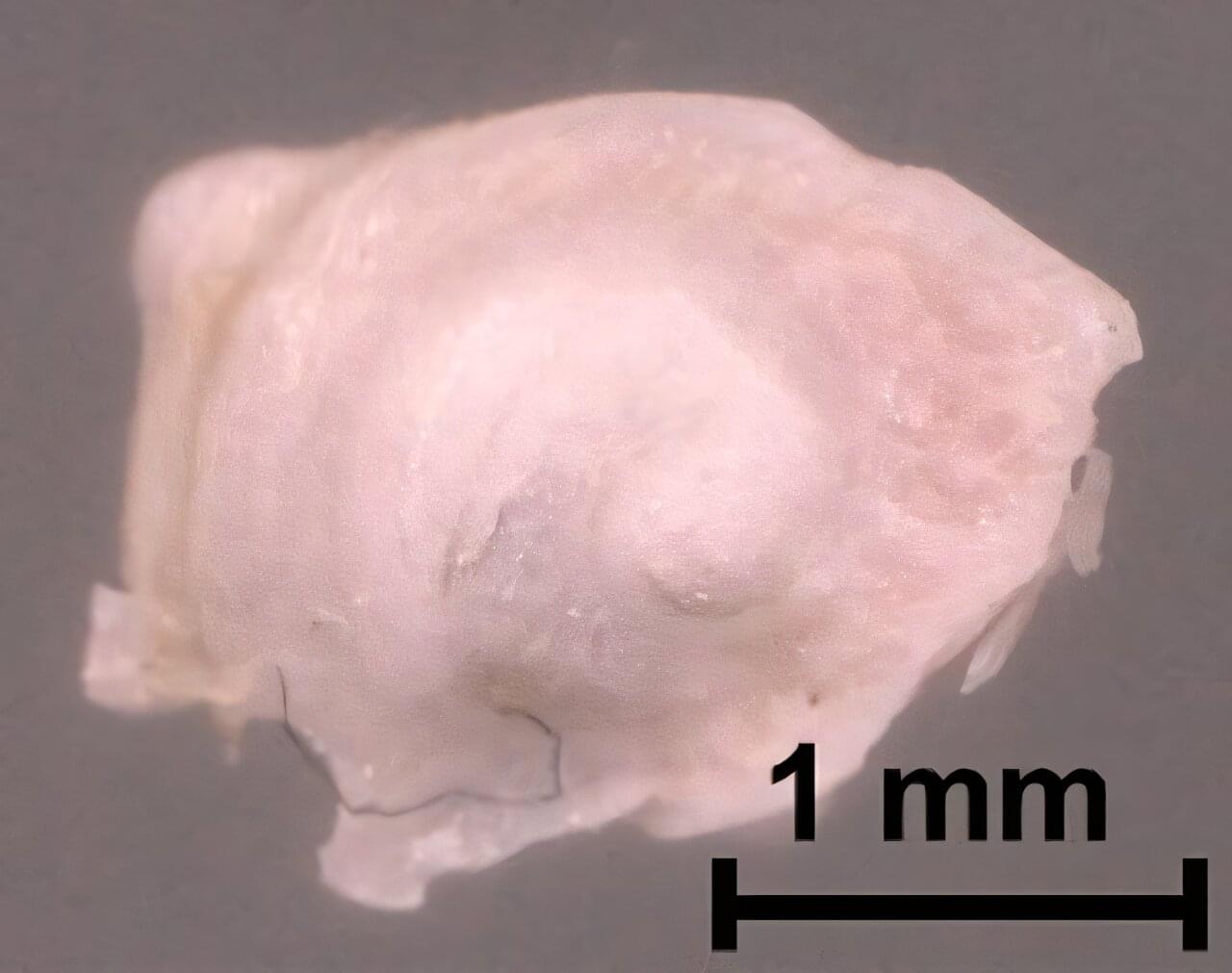
For the first time, researchers have shown that terahertz imaging can be used to visualize internal details of the mouse cochlea with micron-level spatial resolution. The non-invasive method could open new possibilities for diagnosing hearing loss and other ear-related conditions.
“Hearing relies on the cochlea, a spiral-shaped organ in the inner ear that converts sound waves into neural signals,” said research team leader Kazunori Serita from Waseda University in Japan. “Although conventional imaging methods often struggle to visualize this organ’s fine details, our 3D terahertz near-field imaging technique allows us to see small structures inside the cochlea without any damage.”
Terahertz radiation, which falls between microwaves and the mid-infrared region of the electromagnetic spectrum, is ideal for biological imaging because it is low-energy and non-harmful to tissues, scatters less than near-infrared and visible light and can pass through bone while also being sensitive to changes in hydration and cellular structure.
