Toggle light / dark theme

The enzyme telomerase can prevent telomere attrition from happening by extending the length of telomeres. However, in most multicellular organisms, including humans, telomerase expression is switched off, except in germ cells, some types of stem cells, and certain white blood cells. While this might play a role in preventing cancer, as most cancerous cells must switch telomerase expression back on via mutations to enable runaway replication, numerous studies have shown that increasing telomerase through TERT delays aging and increases longevity of model organisms [1].

The small molecule that could

In the lab, this is usually done by introducing genetic vectors carrying a working copy of the gene that codes TERT. It’s this gene that is switched off in somatic cells. However, gene therapies are complex and expensive, and they are just entering the medical mainstream. What if we could do the same using a small molecule?

BOSTON — There currently is no cure for Parkinson’s disease, but scientists may have discovered a potential path to slow or stop the disease. Parkinson’s disease is a brain disorder that can cause movement problems such as tremors, poor balance, and walking difficulties, and it can also impair cognitive function. There is no cure and treatments aim to simply reduce symptoms. But a new study in mice by researchers at Johns Hopkins offers new hope. They found that the interaction of two proteins is key to the buildup of protein clumps in the brain that damage healthy brain cells in Parkinson’s. They also suggest that disrupting these proteins with a drug already approved by the FDA to treat cancer could potentially slow or stop the brain damage associated with Parkinson’s. The next step would be to conduct clinical trials in humans, but that will take time.

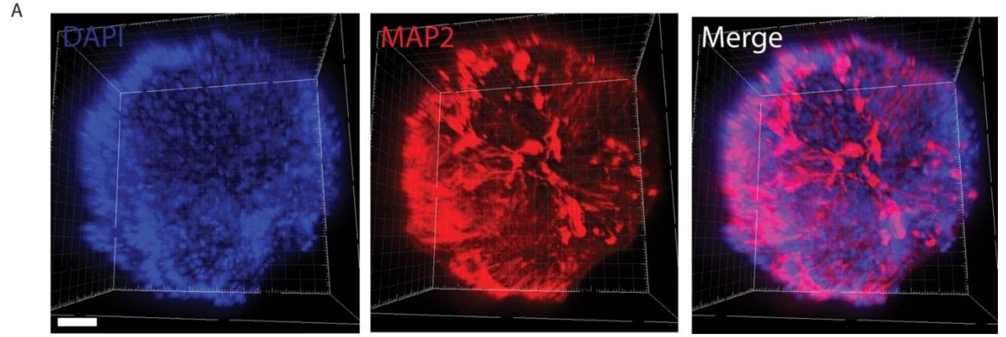
Researchers at University of Michigan have developed a method to produce artificially grown miniature brains—called human brain organoids—free of animal cells that could greatly improve the way neurodegenerative conditions are studied and, eventually, treated.

Over the last decade of researching , scientists have explored the use of as an alternative to mouse models. These self-assembled, 3D tissues derived from embryonic or more closely model the complex structure compared to conventional two-dimensional cultures.

Until now, the engineered network of proteins and molecules that give structure to the cells in , known as extracellular matrices, often used a substance derived from mouse sarcomas called Matrigel. That method suffers significant disadvantages, with a relatively undefined composition and batch-to-batch variability.

Researchers from Johns Hopkins Medicine and the National Institutes of Health’s National Institute on Aging say their study of 40 older adults with obesity and insulin resistance who were randomly assigned to either an intermittent fasting diet or a standard healthy diet approved by the U.S. Department of Agriculture (USDA) offers important clues about the potential benefits of both eating plans on brain health.

Transcription factors (TFs) are proteins that bind to specific DNA sequences, regulating the transcription of genetic information from DNA to messenger RNA (mRNA). These proteins play a pivotal role in the regulation of gene expression, which in turn impacts a wide range of biological processes and brain functions.

The brain is the most complex organ ever created. Its functions are supported by a network of tens of billions of densely packed neurons, with trillions of connections exchanging information and performing calculations. Trying to understand the complexity of the brain can be dizzying. Nevertheless, if we hope to understand how the brain works, we need to be able to map neurons and study how they are wired.

Neuromelanin-sensitive magnetic resonance imaging (NM-MRI) contrast is associated with psychosis severity in antipsychotic-free patients with schizophrenia, according to a study published online Nov. 8 in JAMA Psychiatry.

Kenneth Wengler, Ph.D., from Columbia University in New York City, and colleagues conducted a cross-sectional study involving 42 antipsychotic-free patients with , 53 antipsychotic-free individuals at clinical high risk for psychosis (CHR), and 52 matched healthy controls to replicate previous findings relating NM-MRI, a proxy measure of dopamine function, to psychosis severity. Data were also included for an external validation sample of 16 antipsychotic-naive patients with schizophrenia.

The researchers found that higher Positive and Negative Syndrome Scale positive total scores correlated with higher mean NM-MRI contrast in the psychosis regions of interest (ROI) in the schizophrenia sample.

Schizophrenia, a neurodevelopmental disorder that features psychosis among its symptoms, is thought to arise from disorganization in brain connectivity and functional integration. Now, a recent study in Biological Psychiatry: Cognitive Neuroscience and Neuroimaging, finds differences in functional brain connectivity in people with and without psychosis and schizophrenia that could help researchers understand the neural underpinnings of this disease.

The brain’s cortex is organized in a hierarchical fashion, anchored by the sensorimotor cortex at one end and by multimodal association areas at the other, with the task of integrating incoming sensory information with internal and external sensory signals. The loss of executive control in schizophrenia may stem from disruption of this hierarchical signaling.

Alexander Holmes, a Ph.D. candidate at Monash University who led the study, said, “We used brain imaging and novel mathematical techniques to investigate the hierarchical organization of the brains of individuals with early psychosis and established schizophrenia. This organization is important for brain health, as it regulates how we can effectively respond to and process stimuli from the external world.”