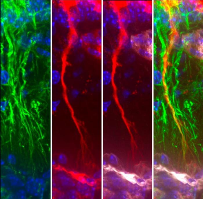
Toggle light / dark theme

Integrated Analysis of Proteomic Marker Databases and Studies Associated with Aging Processes and Age-Dependent Conditions: Optimization Proposals for Biomedical Research

Background: The search for reliable aging biomarkers using proteomic databases and large-scale proteomic studies presents a significant challenge in biogerontology. Existing proteomic databases and studies contain valuable information; however, there is inconsistency in approaches to biomarker selection and data integration. This creates barriers to translating existing knowledge into clinical practice and use in biomedical research. This work analyzed experimental proteomic studies, the content of proteomic databases, and proposed recommendations for optimization and improvement of proteomic database formation and enrichment. Methods: The study utilized publications devoted to proteomic data acquisition methods, proteomic databases, and experimental studies.

Nuclear-lamin-guided plastic positioning and folding of the human genome

Wang et al. systematically depicted the lamins-guided 3D epigenome in human stem cells and demonstrated that lamins not only serve as key regulators of chromatin-NL tethering and large-scale genome organization but also are essential for the spatial positioning and clustering of nuclear speckle through direct interactions within the nuclear interior.

How the microbiome and a fiber-rich diet help fight melanoma

Scientists at the Peter Doherty Institute for Infection and Immunity (Doherty Institute) have uncovered how the gut microbiota help the immune system fight melanoma, explaining why patients with a fiber-rich diet and balanced gut bacteria tend to respond better to cancer immunotherapies.

The study, published in Immunity, shows that molecules produced by upon digestion of dietary fiber can improve the function of cancer-fighting . The research team, led by the Doherty Institute, in collaboration with the Peter MacCallum Cancer Center (Peter Mac) and Monash University, found that these digestive by-products influence melanoma progression by naturally boosting killer T cell function in pre-clinical cancer models.

Improved method offers broader, faster detection of protein-ligand interactions

EMBL scientists have improved a protein analysis technique, significantly expanding its use and making it 100 times faster.

Swedish chemist Jöns Jacob Berzelius, in a letter to a fellow chemist, first suggested the name “proteins” for a particular class of biological substances, deriving it from the Greek word proteios, meaning “primary” or “of first importance.” Although scientists in the 1830s knew very little about proteins, it was already clear how essential they were to living organisms.

Long-known as the “workhorses of the cell,” proteins are responsible for powering nearly every function in the body. Often critical to this is their interactions with other small molecules known as ligands. In a new study published in Nature Structural and Molecular Biology, EMBL researchers introduce HT-PELSA, a high-throughput adaptation of an earlier tool that detects these interactions. This new tool can process samples at an unprecedented scale, a breakthrough that promises to accelerate and our understanding of fundamental biological processes.

Research reveals shared genetic roots for psychiatric and neurological disorders

Researchers from the Center for Precision Psychiatry at the University of Oslo and Oslo University Hospital have discovered extensive genetic links between neurological disorders like migraine, stroke and epilepsy, and psychiatric illnesses such as schizophrenia and depression. Published in Nature Neuroscience, this research challenges longstanding boundaries between neurology and psychiatry and points to the need for more integrated approaches to brain disorders.

“We found that psychiatric and neurological disorders share to a greater extent than previously recognized. This suggests that they may partly arise from the same underlying biology, contrasting the traditional view that they are separate disease entities. Importantly, the genetic risk was closely linked to brain biology,” states Olav Bjerkehagen Smeland, psychiatrist and first author.

Nonsurgical treatment shows promise for targeted seizure control

Rice University bioengineers have demonstrated a nonsurgical way to quiet a seizure-relevant brain circuit in an animal model. The team used low-intensity focused ultrasound to briefly open the blood-brain barrier (BBB) in the hippocampus, delivered an engineered gene therapy only to that region and later flipped an on-demand “dimmer switch” with an oral drug.

The research shows that a one-time, targeted procedure can modulate a specific brain region without impacting off-target areas of the brain. It is published in and featured on the cover of ACS Chemical Neuroscience.

“Many are driven by hyperactive cells at a particular location in the brain,” said study lead Jerzy Szablowski, assistant professor of bioengineering and a member of the Rice Neuroengineering Initiative. “Our approach aims the therapy where it is needed and lets you control it when you need it, without surgery and without a permanent implant.”

It’s not just in your head: Stress may lead to altered blood flow in the brain

While the exact causes of neurodegenerative brain diseases like Alzheimer’s and dementia are still largely unknown, researchers have been able to identify a key characteristic in affected brains: reduced blood flow. Building upon this foundational understanding, a team at Penn State recently found that a rare neuron that is extremely vulnerable to anxiety-induced stress appears to be responsible for regulating blood flow and coordinating neural activity in mice.

The researchers found that eliminating type-one nNOS neurons—which make up less than 1% of the brain’s 80 billion neurons and die off when exposed to too much stress—resulted in a drop in both blood flow and in mice’s brains, demonstrating the impact this neuron type has on the proper brain functions of animals, including humans.

The research appears in eLife.

Angstrom-level imaging and 2D surfaces allow real-time tracking and steering of DNA

Pictures of DNA often look very tidy—the strands of the double helix neatly wind around each other, making it seem like studying genetics should be relatively straightforward. In truth, these strands aren’t often so perfectly picturesque. They are constantly twisting, bending, and even being repaired by minuscule proteins. These are movements on the nanoscale, and capturing them for study is extremely challenging. Not only do they wriggle about, but the camera’s fidelity must be high enough to focus on the tiniest details.

Researchers from the University of Illinois Urbana-Champaign (U. of I.) have been working on resolving a grand challenge for , and more specifically, : how to take a high-resolution image of DNA to facilitate study.

Using a number of compute resources, including NCSA’s Delta, Aleksei Aksimentiev, a professor of physics at U. of I, and Dr. Kush Coshic, formerly a graduate research assistant in the Center for Biophysics and Quantitative Biology and the Beckman Institute for Advanced Science and Technology at U. of I., and currently a postdoctoral fellow at the Max Planck Institute of Biophysics, recently made significant contributions to solving this challenge. They did it by focusing on two specific problems: creating a “camera” that could capture the molecular movement of DNA, and by creating an environment in which they could predictably direct the movement of the DNA strands.

Nanorobots guide stem cells to become bone cells via precise pressure

For the first time, researchers at the Technical University of Munich (TUM) have succeeded in using nanorobots to stimulate stem cells with such precision that they are reliably transformed into bone cells. To achieve this, the robots exert external pressure on specific points in the cell wall. The new method offers opportunities for faster treatments in the future.

Prof. Berna Özkale Edelmann’s nanorobots consist of tiny gold rods and plastic chains. Several million of them are contained in a gel cushion measuring just 60 micrometers, together with a few . Powered and controlled by , the robots, which look like tiny balls, mechanically stimulate the cells by exerting pressure.

“We heat the gel locally and use our system to precisely determine the forces with which the nanorobots press on the cell—thereby stimulating it,” explains the professor of nano-and microrobotics at TUM. This mechanical stimulation triggers biochemical processes in the cell. Ion channels change their properties, and proteins are activated, including one that is particularly important for bone formation.

/* */