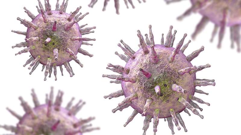
Toggle light / dark theme

In a recent study published in Nature Communications, a team of researchers at the Carl R. Woese Institute for Genomic Biology reports a new, robust computational toolset to extract biological relationships from large transcriptomics datasets. These efforts will help scientists better investigate cellular processes.

Living organisms are governed by their genome—an instruction manual written in the language of DNA that dictates how an organism grows, survives, and reproduces. By regulating the abundance of different RNA transcripts, cells control their protein expression level, thereby shaping their functions and responses to the environment.

Transcriptomics is the study of gene expression through cataloging the presence and abundance levels of active RNA transcripts generated from the genome under different conditions. Through the lens of RNA, transcriptomics technologies allow scientists to study the that enable life and cause disease, as well as assess the biological effects of therapeutics.

Unlocking Real-Time Inflammation Monitoring with Active-Reset Protein Sensors.

Imagine a tiny device that could continuously monitor your body’s inflammation levels, offering insights in real time to help manage diseases. While sensors for small molecules like glucose have existed for years, tracking proteins—a critical component in understanding inflammation—has been a major challenge. Proteins are present at much lower levels in the body, and traditional sensors struggle with slow response times due to their high-affinity binding to these molecules.

A team led by Zargartalebi has now overcome this barrier by introducing active-reset protein sensors. These sensors employ high-frequency oscillations of positive voltage to rapidly release bound protein molecules from their sensing electrodes. This breakthrough allows the sensors to reset in under a minute, enabling continuous tracking of protein levels.

The researchers incorporated these sensors into a microneedle device, which was successfully tested in mice to monitor cytokines—key markers of inflammation associated with diabetes. Unlike previous approaches, this method is not only simple to implement but can also be adapted to various sensor designs, potentially revolutionizing how we monitor and respond to conditions like chronic inflammation.

By bringing real-time protein monitoring closer to reality, active-reset sensors could pave the way for more dynamic and responsive healthcare, ensuring better disease management and prevention.

[#biosensors](https://www.facebook.com/hashtag/biosensors?__eep__=6&__cft__[0]=AZXpWTjqoXcElrcVMft-YBHF33SrUIfWG14SxebDv6jc7ayB9obklbDJ5BXDEcXMFhRK4gARwyDe24ko0Pjq0q5uSGpyaiia1YlxceB88HQ4AkLMRRKBaP_TJR0TQWWUQlYvZFrxBhxODGJhyWYo_RjJbdkcNE9A_kXuT2XJxXfpKD1LGhewSjGBaGndlInALDM&__tn__=*NK-R) [#invivo](https://www.facebook.com/hashtag/invivo?__eep__=6&__cft__[0]=AZXpWTjqoXcElrcVMft-YBHF33SrUIfWG14SxebDv6jc7ayB9obklbDJ5BXDEcXMFhRK4gARwyDe24ko0Pjq0q5uSGpyaiia1YlxceB88HQ4AkLMRRKBaP_TJR0TQWWUQlYvZFrxBhxODGJhyWYo_RjJbdkcNE9A_kXuT2XJxXfpKD1LGhewSjGBaGndlInALDM&__tn__=*NK-R) [#sensing](https://www.facebook.com/hashtag/sensing?__eep__=6&__cft__[0]=AZXpWTjqoXcElrcVMft-YBHF33SrUIfWG14SxebDv6jc7ayB9obklbDJ5BXDEcXMFhRK4gARwyDe24ko0Pjq0q5uSGpyaiia1YlxceB88HQ4AkLMRRKBaP_TJR0TQWWUQlYvZFrxBhxODGJhyWYo_RjJbdkcNE9A_kXuT2XJxXfpKD1LGhewSjGBaGndlInALDM&__tn__=*NK-R) [#inflammation](https://www.facebook.com/hashtag/inflammation?__eep__=6&__cft__[0]=AZXpWTjqoXcElrcVMft-YBHF33SrUIfWG14SxebDv6jc7ayB9obklbDJ5BXDEcXMFhRK4gARwyDe24ko0Pjq0q5uSGpyaiia1YlxceB88HQ4AkLMRRKBaP_TJR0TQWWUQlYvZFrxBhxODGJhyWYo_RjJbdkcNE9A_kXuT2XJxXfpKD1LGhewSjGBaGndlInALDM&__tn__=*NK-R) [#Microneedle](https://www.facebook.com/hashtag/microneedle?__eep__=6&__cft__[0]=AZXpWTjqoXcElrcVMft-YBHF33SrUIfWG14SxebDv6jc7ayB9obklbDJ5BXDEcXMFhRK4gARwyDe24ko0Pjq0q5uSGpyaiia1YlxceB88HQ4AkLMRRKBaP_TJR0TQWWUQlYvZFrxBhxODGJhyWYo_RjJbdkcNE9A_kXuT2XJxXfpKD1LGhewSjGBaGndlInALDM&__tn__=*NK-R)

[#regenerativesensors](https://www.facebook.com/hashtag/regenerativesensors?__eep__=6&__cft__[0]=AZXpWTjqoXcElrcVMft-YBHF33SrUIfWG14SxebDv6jc7ayB9obklbDJ5BXDEcXMFhRK4gARwyDe24ko0Pjq0q5uSGpyaiia1YlxceB88HQ4AkLMRRKBaP_TJR0TQWWUQlYvZFrxBhxODGJhyWYo_RjJbdkcNE9A_kXuT2XJxXfpKD1LGhewSjGBaGndlInALDM&__tn__=*NK-R) [#pointofcare](https://www.facebook.com/hashtag/pointofcare?__eep__=6&__cft__[0]=AZXpWTjqoXcElrcVMft-YBHF33SrUIfWG14SxebDv6jc7ayB9obklbDJ5BXDEcXMFhRK4gARwyDe24ko0Pjq0q5uSGpyaiia1YlxceB88HQ4AkLMRRKBaP_TJR0TQWWUQlYvZFrxBhxODGJhyWYo_RjJbdkcNE9A_kXuT2XJxXfpKD1LGhewSjGBaGndlInALDM&__tn__=*NK-R) [#diagnostics](https://www.facebook.com/hashtag/diagnostics?__eep__=6&__cft__[0]=AZXpWTjqoXcElrcVMft-YBHF33SrUIfWG14SxebDv6jc7ayB9obklbDJ5BXDEcXMFhRK4gARwyDe24ko0Pjq0q5uSGpyaiia1YlxceB88HQ4AkLMRRKBaP_TJR0TQWWUQlYvZFrxBhxODGJhyWYo_RjJbdkcNE9A_kXuT2XJxXfpKD1LGhewSjGBaGndlInALDM&__tn__=*NK-R) [#cytokines](https://www.facebook.com/hashtag/cytokines?__eep__=6&__cft__[0]=AZXpWTjqoXcElrcVMft-YBHF33SrUIfWG14SxebDv6jc7ayB9obklbDJ5BXDEcXMFhRK4gARwyDe24ko0Pjq0q5uSGpyaiia1YlxceB88HQ4AkLMRRKBaP_TJR0TQWWUQlYvZFrxBhxODGJhyWYo_RjJbdkcNE9A_kXuT2XJxXfpKD1LGhewSjGBaGndlInALDM&__tn__=*NK-R)

Ingestible devices are often used to study and treat hard-to-reach tissues in the body. Swallowed in pill form, these capsules can pass through the digestive tract, snapping photos or delivering drugs.

While in their simplest form, these devices are passively transported through the gut, there are a wide range of applications where you may want a device to attach to the tissue or other flexible materials. A rich history of biologically inspired solutions exist to address this need, ranging from cocklebur-inspired Velcro to slug-inspired medical adhesives, but the creation of on-demand and reversible attachment mechanisms that can be incorporated into millimeter-scale devices for biomedical sensing and diagnostics remains a challenge.

A new interdisciplinary effort led by Robert Wood, the Harry Lewis and Marlyn McGrath Professor of Engineering and Applied Sciences in the Harvard John A. Paulson School of Engineering and Applied Sciences (SEAS), and James Weaver, of Harvard’s Wyss Institute, has drawn inspiration from an unexpected source: the world of parasites.

Proteins are so much more than nutrients in food. Virtually every reaction in the body that makes life possible involves this large group of molecules. And when things go wrong in our health, proteins are usually part of the problem.

In certain types of heart disease, for instance, the proteins in cardiac tissue, seen with , are visibly disordered. Alex Dunn, professor of chemical engineering, describes proteins like the beams of a house: “We can see that in unhealthy heart muscle cells, all of those beams are out of place.”

Proteins are the workhorses of the cell, making the biochemical processes of life possible. These workhorses include enzymes, which bind to other molecules to speed up reactions, and antibodies that attach to viruses and prevent them from infecting cells.

Researchers have used a mouse model to show that infections in the intestine can change the composition of bile, a fluid that is generated in the liver and is crucial to digestion. Bile aids in the absorption of fat and contributes to defense against infections. This study has suggested that intestinal infections can alter microbes in the gastrointestinal tract, or the microbiome, and modify the immune system. Although the work was conducted in mice, the researchers suggested that their conclusions also apply to humans. The findings have been reported in Nature Microbiology.

“The changes we detected in the composition of bile with infection are beneficial for the intestine to clear infection,” said corresponding study author Matthew Waldor, MD, PhD, of Brigham and Women’s Hospital. “Our findings reveal the intricate and dynamic nature of bile composition, shedding new light on the liver’s critical role in defending the intestine from infection. These insights enhance our understanding of the liver’s broader functions in regulating physiological stability and metabolic processes.”

Yale researchers have made an unexpected discovery—turncoat T cells that help a tumor evade other cancer-fighting immune T cells—in a study of patients living with advanced melanoma.

The study by Yale Cancer Center (YCC) researchers at Yale School of Medicine (YSM) discovered that not all CD8+ T cells are allies in a body’s fight against . Patients living with severe who had increased levels of suppressor, regulatory CD8+ T cells had worse survival outcomes.

The study is published in the journal Nature Immunology.

Death might seem like a pure loss, the disappearance of what makes a living thing distinct from everything else on our planet. But zoom in closer, to the cellular level, and it takes on a different, more nuanced meaning. There is a challenge in simply defining what makes an individual cell alive or dead. Scientists today are working to understand the various ways and reasons that cells disappear, and what these processes mean to biological systems. In this episode, cellular biologist Shai Shaham talks to Steven Strogatz about the different forms of cell death, their roles in evolution and disease, and why the right kinds and patterns of cell death are essential to our development and well-being.

Listen on Apple Podcasts, Spotify, TuneIn or your favorite podcasting app, or you can stream it from Quanta.

Improvements in cancer prevention and screening have averted more deaths from five cancer types combined over the past 45 years than treatment advances, according to a modeling study led by researchers at the National Institutes of Health (NIH).

The study, published Dec. 5, 2024, in JAMA Oncology, looked at deaths from breast, cervical, colorectal, lung, and prostate cancer that were averted by the combination of prevention, , and advances.

The researchers focused on these five cancers because they are among the most common causes of cancer deaths and strategies exist for their prevention, early detection, and/or treatment. In recent years, these five cancers have made up nearly half of all new cancer diagnoses and deaths.