Wearable technologies are starting to reshape how people manage health. Continuous glucose monitors that measure blood sugar levels in diabetes patients have already shown the power of tracking an important molecule in real time. The next leap is to track other medically important molecules. However, doing so is far more difficult because most of those molecules are present at much lower concentrations than glucose.
One area such wearable technologies could transform is drug therapy. Many powerful medications are still managed through blood tests that offer only occasional snapshots of how a patient’s body is processing treatment. For drugs that must be dosed precisely to avoid harm, clinicians can miss the point at which dosing becomes ineffective or begins to threaten the organs responsible for processing the drug.
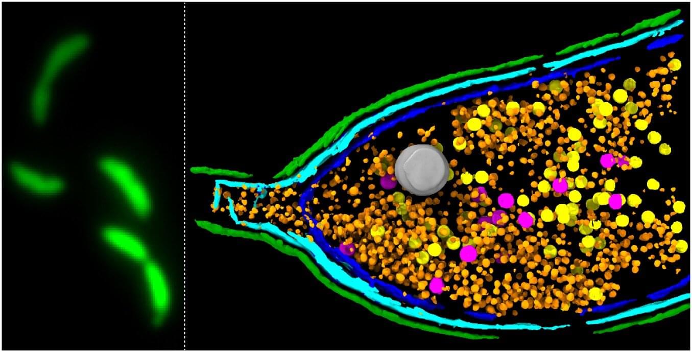
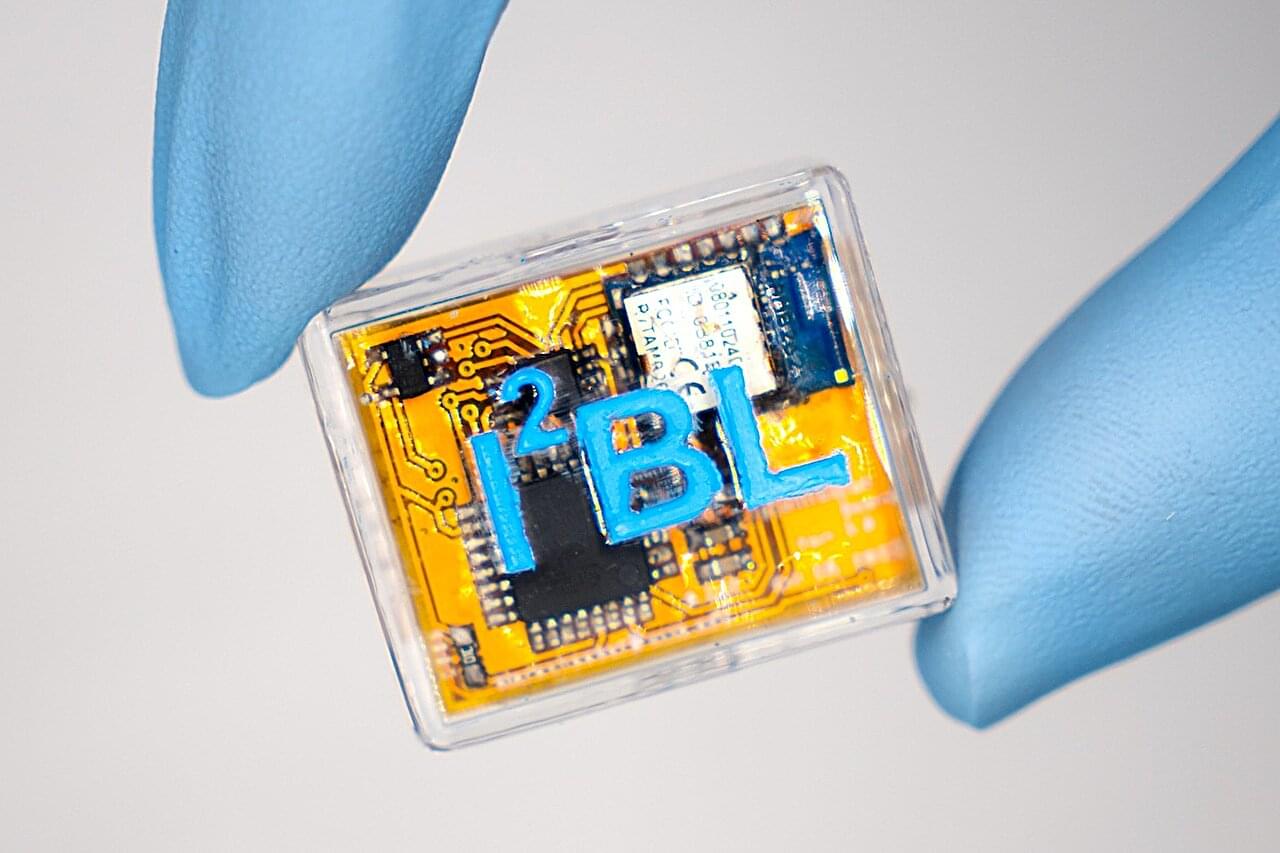
A UCLA-led research team has now developed a microneedle sensor platform designed to address that problem through continuous, minimally invasive monitoring in skin. In a study published in Science Translational Medicine, the researchers showed in rats that the sensors could operate continuously for six days, track drug concentrations over time and provide insight into kidney and liver function by measuring how quickly the body cleared those drugs.

