Among patients with newly diagnosed metastatic BreastCancer (MBC), a clinical cohort trial evaluated early treatment response using 18F-fluorodeoxyglucose positron emission tomography (FDG-PET) compared with standard computed tomography (CT) assessment.
FDG-PET after only 2 weeks of treatment identified patients with MBC with distinct long-term outcomes. Incorporating early FDG-PET can improve outcome estimation of standard CT assessment.
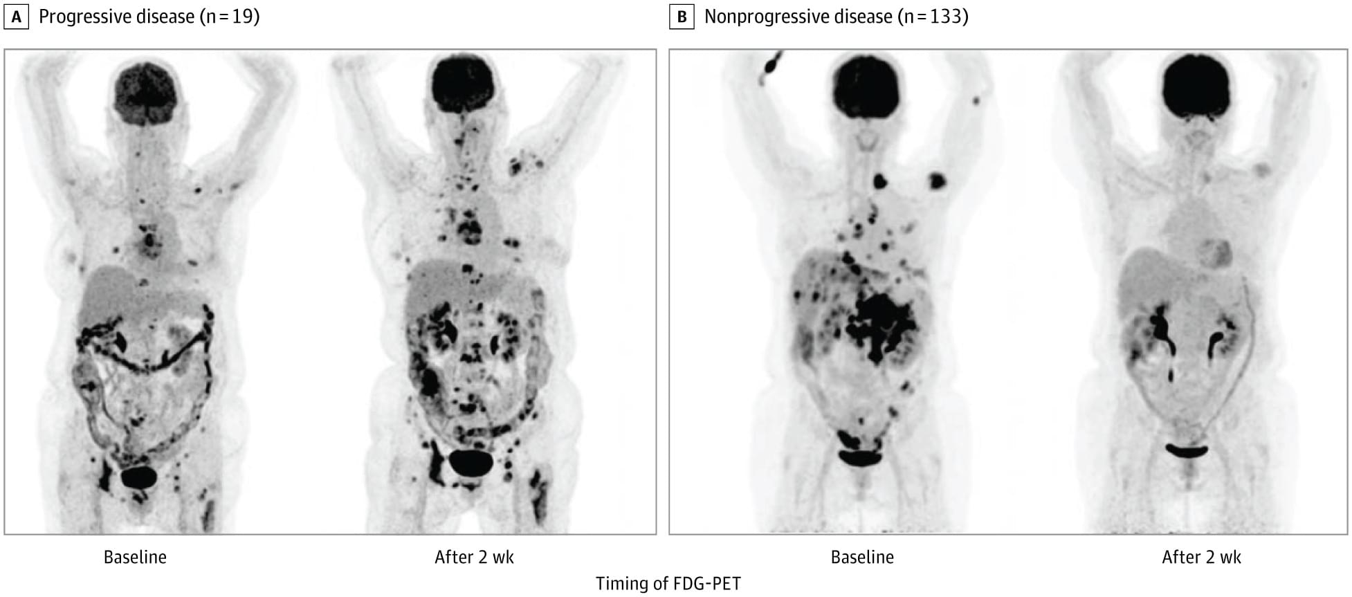
Question Does repeated 18 F-fluorodeoxyglucose positron emission tomography (FDG-PET) after 2 weeks of treatment improve outcome estimation in metastatic breast cancer compared to standard diagnostics?
Findings This clinical cohort trial including 200 patients found that those without disease progression on early FDG-PET had better median progression-free survival (PFS) and overall survival (OS) than patients with disease progression. Among patients without progression on computed tomography (CT) after 8 weeks, those with progression on early FDG-PET had a median OS of 22.3 months, whereas patients without progression on both CT and early FDG-PET had an OS of 50.1 months.
Meaning Early FDG-PET imaging may improve outcome estimation in newly diagnosed metastatic breast cancer compared to standard CT alone.