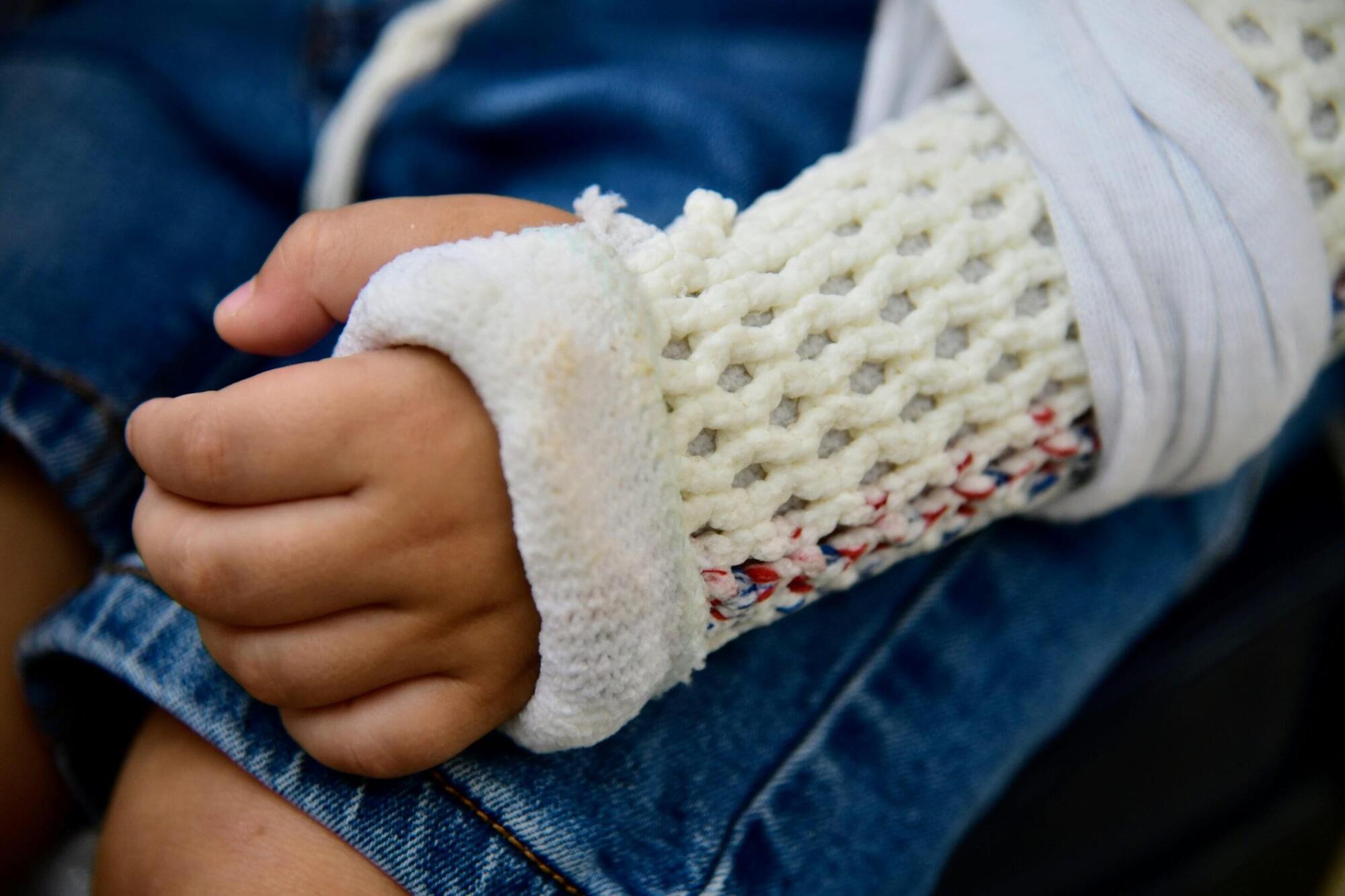
Toggle light / dark theme

Cutting CAR T Costs via Decentralization, Vector-Alternatives, and Artificial Intelligence

High manufacturing costs are limiting patient access to CAR T cell therapies, according to new research, which indicates that decentralization, vector-free modification technologies, and AI would help make production cheaper.

Making CAR T therapies is an expensive business. A recent study suggested that producing a single batch can cost anywhere between $170,000 and $220,000, depending on the logistical, processing, and distribution steps involved.

The fundamental problem is that CAR T production is not a good fit for centralized manufacturing, according to Martin Bonamino, PhD, leader of the experimental cancer immunotherapy group at Brazil’s National Cancer Institute (INCA).

Emerging role of mitochondrial calcium levels in cellular senescence and in switching cell fates

Manipulating senescent cells by eliminating them or by modifying their activity has attracted huge interest for its potential to delay or even treat many age-related diseases, and to improve healthy aging. Mitochondria, and in particular their calcium levels, have emerged as key regulators of cellular senescence, cell death and the balance between the two, and might constitute targets for novel strategies to stifle the viability or properties of senescent cells.

Conserved brain-wide emergence of emotional response from sensory experience in humans and mice

Emotional responses to sensory experience are central to the human condition in health and disease. We hypothesized that principles governing the emergence of emotion from sensation might be discoverable through their conservation across the mammalian lineage. We therefore designed a cross-species neural activity screen, applicable to humans and mice, combining precise affective behavioral measurements, clinical medication administration, and brain-wide intracranial electrophysiology. This screen revealed conserved biphasic dynamics in which emotionally salient sensory signals are swiftly broadcast throughout the brain and followed by a characteristic persistent activity pattern. Medication-based interventions that selectively blocked persistent dynamics while preserving fast broadcast selectively inhibited emotional responses in humans and mice.

Atomic-level view of plant cell death enzyme offers path to safer crop protection

In a discovery three decades in the making, scientists at Rutgers and Brookhaven National Laboratory have acquired detailed knowledge about the internal structures and mode of regulation for a specialized protein and are proceeding to develop tools that can capitalize on its ability to help plants combat a wide range of diseases.

The work, which exploits a natural process where plant cells die on purpose to help the host plant stay healthy, is expected to have wide applications in the agricultural sector, offering new ways to protect major food crops from a variety of devastating diseases, the scientists said.

In a study published in Nature Communications, a team led by Eric Lam at Rutgers University-New Brunswick and Qun Liu at Brookhaven National Laboratory in New York reported that advanced crystallography and computer modeling techniques have enabled them to obtain the best picture yet of a pivotal plant protease, a that cuts other proteins, known as metacaspase 9.

3D genome mapping technology sheds light on how plants regulate photosynthesis

Chinese researchers have developed a technology that sheds light on how the three-dimensional (3D) organization of plant genomes influences gene expression—especially in photosynthesis.

The research, which was led by Prof. Xiao Jun at the Institute of Genetics and Developmental Biology of the Chinese Academy of Sciences, in collaboration with BGI Research, is published in Science Advances.

The innovative method not only provides a more precise tool for understanding the intricate 3D interactions between genes, but also highlights the critical role of long-range chromatin interactions in .

Custom-designed polymers open new path to electrochemical separations for sustainable drug manufacturing

Enantiomers, or molecule pairs that are mirror images of each other, make up more than half of FDA-approved drugs in use today, including those used in treatments for cancer, neurologic diseases and arthritis. Separating enantiomers is critical for drug manufacturing because the effect of each molecule in the pair can be very different—for example, one enantiomer might cure a headache while its mirror-image could cause a headache.

Faster and more accurate separations would help with the overall drug discovery and screening process, but by their very nature, enantiomers—which have identical compositions and only differ by not being superimposable (think left hand and right hand)—are notoriously difficult to separate.

An effort by a group of researchers at the University of Illinois Urbana-Champaign to find an efficient, sustainable way to perform these critical enantiomer separations is the focus of a new study published in the Journal of the American Chemical Society.

Protein misfolding and aggregation in Alzheimer’s disease and Type 2 Diabetes Mellitus

In a diseased condition, most of the time, target proteins attain toxicity following their transition from a α-helix to a β-sheet form [18]. Although numerous functional native proteins possess β-sheet conformations within them, the transition from an α-helix to a β-sheet is characteristic of amyloid deposits [19], and often associated with the change of a physiological function to a pathological one. Such abnormal conformational transition exposes hydrophobic amino acid residues and promotes protein aggregation [18, 20]. The toxic proteins often interact with other native proteins and may catalyze their transition into a toxic sate, and hence they are called infective conformations [18]. The newly formed toxic proteins can repeat this cycle to intiate a self-sustaining loop; thereby amplifying the toxicity to generate a catastrophic effect, beyond homeostatic reparative mechanisms, to eventually impair cellular function or induce cellular demise [21].

Proteins function properly when their constituent amino acids fold correctly [22]. On the other hand, misfolded proteins assemble into insoluble aggregates with other proteins and can be toxic for the cells [18, 20]. Ataxin-1 is highly prone to misfolding due to inherited gene defects that cause neurodegenerative diseases (NDDs), which is mainly due the repetition of glutamine within its amino acid chain; the toxicity of this protein being directly proportional to the number of glutamines [23]. There are 21 proteins that mainly interact with ataxin-1 and influence its folding or misfolding, 12 of which increase the toxicity of ataxin-1 for nerve cells, while 9 of the identified proteins reduce its toxicity [23]. Ataxin-1 resembles a double twisted spiral or helix and has a special structure, termed a “coiled coil domain”, that promotes aggregation. Proteins which possess “coiled coil domain” and interact with ataxin-1 have been reported to enhance promotion of ataxin-1 aggregation and toxic effects [24].

The gradual accumulation of misfolded proteins in the absence of their appropriate clearance can cause amyloid disease, the most prevalent one being AD. Parkinson’s disease and Huntington’s disease have similar amyloid origins [25]. These diseases can be sporadic or familial and their incidence increases dramatically with age. The mechanistic explanation for this correlation is that as we age (and are subjected to increasing numbers of mutations and/or oxidative stress causing changes to protein structure, etc.), the delicate balance of the synthesis, folding, and degradation of proteins is disturbed, ensuing in the production, accumulation and aggregation of misfolded proteins [26].

/* */