Toggle light / dark theme

When “Artificial Neurons” Can Talk Directly to the “Brain”

*** This content was analyzed and written by AI for informational purposes only.
*** Please consult a specialist for professional advice.

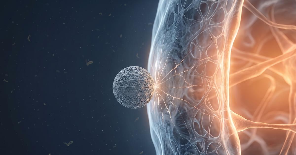
The world is entering an era where “technology” and “living organisms” merge into one. Most recently, in 2026, a research team from Northwestern University created a landmark breakthrough by developing “Printed Neurons.” These are not designed just to mimic biology—they can actually “transmit signals” to communicate with living brain cells!

Why is this a big deal?
Typically, the silicon-based computers we use today operate entirely differently from the human brain. Computers consume massive amounts of power and are rigid. In contrast, our brains use only about 20 watts (less than some lightbulbs) and are incredibly flexible.
Creating artificial neurons that “speak the same language as the brain” is the key to treating diseases that were once considered incurable.

Innovations in “Electronic Ink” and “3D Printing“
At the heart of this research lies a leap forward in materials science and engineering:
• Nanomaterials (MoS₂ and Graphene): Researchers used these materials to create a specialized “ink” for printing neural networks. These materials are unique for being both flexible and excellent conductors of electricity.
• Aerosol Jet Printing: This technology allows for nano-level precision printing on flexible plastic sheets, designed to contour perfectly to human tissue.
• Biomimicry: These artificial cells can generate electrical signals called “Spikes,” matching the rhythm and speed of actual biological neurons.

Proven! Successful Communication with a “Mouse Brain“
The research team tested the connection between these printed neurons and mouse brain tissue. The results showed that the mouse brain cells could receive and respond to signals from the artificial device as if they were from their own kind. This is vital evidence that humans can create devices that interface seamlessly with the nervous system.

Printed Artificial Neurons Connect With Real Brain Cells | WION News

Northwestern University engineers developed flexible, printed artificial neurons that communicate directly with living brain cells, marking a major breakthrough for brain-machine interfaces and neuroprosthetics. Using aerosol jet printing with materials like molybdenum disulfide, these devices generate signals that trigger responses in mouse brain tissue.

#neurons #braincells #wion.

About Channel:

WION The World is One News examines global issues with in-depth analysis. We provide much more than the news of the day. Our aim is to empower people to explore their world. With our Global headquarters in New Delhi, we bring you news on the hour, by the hour. We deliver information that is not biased. We are journalists who are neutral to the core and non-partisan when it comes to world politics. People are tired of biased reportage and we stand for a globalized united world. So for us, the World is truly One.

Please keep discussions on this channel clean and respectful and refrain from using racist or sexist slurs and personal insults.

Check out our website: http://www.wionews.com.

We Are Creating Artificial Brains

The Big Why explores the cutting edge of science and technology: Artificial Brains! 🧠🤖 In this mind-blowing video, we dive into the quest to replicate the human brain’s complexity and power in a machine.

Discover the various approaches scientists are taking, from simulating neural networks to building brain-like hardware. We’ll examine the potential of this technology to revolutionize medicine, robotics, and even our understanding of consciousness.

But we won’t shy away from the big questions either: Could artificial brains surpass human intelligence? What are the ethical implications of creating conscious machines? Join us as we ponder the future of AI and the potential for a technological singularity.

#ArtificialBrain #AI #Neuroscience #Robotics #FutureTech #Consciousness #Singularity #thebigwhy

00:00 — Intro
01:33 — Overview
03:39 — Approaches to Brain Simulation
06:02 — Artificial Brain Thought Experiment
07:40 — Outro.

Glial Cells as Emerging Therapeutic Targets in Neurodegenerative Diseases: Mechanistic Insights and Translational Perspectives

Neurodegenerative diseases such as Alzheimer’s disease (AD), Parkinson’s disease (PD), Huntington’s disease, multiple sclerosis, and amyotrophic lateral sclerosis share converging mechanisms of neuronal dysfunction, including protein aggregation, oxidative stress, and chronic neuroinflammation. Glial cells, once considered passive supporters, are now recognized as central drivers of these processes, offering both pathogenic triggers and therapeutic opportunities. Yet, despite compelling preclinical evidence, the translation of glial-targeted therapies into clinical success has been limited. This review provides a critical synthesis of current knowledge by examining therapeutic strategies through the lens of their translational challenges and failures.

Insular input to the prelimbic cortex underlies social affective behavior in rats

Delayed onset of hemichorea and dystonia can occur after cortical temporal Stroke, even when imaging reveals small acute ischemic lesions and routine blood tests are normal.


This case report describes delayed hemichorea-dystonia following right carotid endarterectomy for proximal internal carotid artery stenosis in a 79-year-old man.

Genetic Landscape and Diagnostic Outcomes of UK Patients With Congenital Myopathies and Muscular Dystrophies Over a 10-Year Period

This website uses a security service to protect against malicious bots. This page is displayed while the website verifies you are not a bot.

White matter injury may lead to neurodegeneration

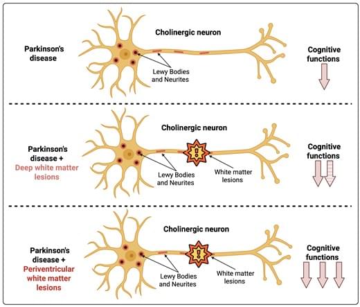
The brain is equally divided into grey and white matter. Grey matter contains the brain’s processing hubs, linked by an information highway — the white matter. Although white matter damage is a defining feature of multiple sclerosis and is also seen in neurodegeneration including Alzheimer’s and Parkinson’s disease, the consequences of white matter damage are not well understood.

The team created localised damage to myelin – the main component of white matter – in a well-defined brain circuit and followed what happened over time. They found that small, localised myelin damage triggered a striking response in a connected, remote grey matter region. Neuronal activity fell, microglia – the brain’s immune cells – became activated, and connections between neurons were lost.

Crucially, these changes were not permanent. After myelin was regenerated, neuronal activity recovered, connections between neurons returned, and the inflammatory response subsided.

The study also challenges a common assumption about brain inflammation. Grey matter inflammation is traditionally viewed as harmful. But here, the team found that this transient response was part of the repair process itself. When they prevented grey matter inflammation, myelin regeneration was impaired.

Conversely, when the team blocked myelin regeneration, the grey matter response did not resolve and instead became chronic. This suggests that failed myelin regeneration may help drive the persistent low-grade inflammation seen in neurodegenerative disease. ScienceMission sciencenewshighlights.


Damage to white matter in the brain can trigger features associated with neurodegenerative disease, The researchers have discovered in a new study published in the journal Nature.

The role of noradrenergic innervation and β-cell dedifferentiation in diabetes

Noradrenergic innervation and β-cell dedifferentiation in diabetes.

Dedifferentiation, a survival mechanism whereby mature β-cells revert to a nonfunctional state under metabolic stress, represents a fundamental driver of β-cell failure in type 2 diabetes.

Dedifferentiation is reversible, primarily through dietary intervention or bariatric surgery, and redifferentiation may promote type 2 diabetes remission.

Noradrenergic fiber density is increased in diabetic pancreases and correlates with β-cell dedifferentiation, suggesting that altered signaling may trigger the process.

A link between diet, redifferentiation, reduction of noradrenergic fibers, and type 2 diabetes remission has been hypothesized.

The review proposes that targeting pancreatic noradrenergic innervation could be a novel therapeutic strategy to reverse β-cell dedifferentiation, restore insulin function, and achieve type 2 diabetes remission. sciencenewshighlights ScienceMission https://sciencemission.com/noradrenergic-innervation–in-diabetes


Baicalein Alleviates Iron Overload-Induced Ferroptosis and Osteogenic Blockade in Osteoblasts by Activating the Nrf2/GPX4 Pathway

JUST PUBLISHED:Click here to read the latest free, Open Access article from BMEF.


The transcription factor Nrf2 orchestrates cellular defenses against redox imbalance and lipid peroxidation, partly through regulating the expression of 2 key gatekeepers of ferroptosis: SLC7A11 and GPX4 [44]. As such, the Keap1/Nrf2 pathway is recognized as a master regulator of ferroptosis in osteoblasts [45]. Under stress conditions, Nrf2 dissociates from the Keap1–Nrf2 complex, translocates into the nucleus, and initiates the transcription of genes containing antioxidant response elements [46]. Previous studies have reported that Nrf2 activation protects osteoblasts from ferroptosis in bone tissue and alleviates osteoporosis [28,47]. Consistently, we observed that under iron overload conditions, baicalein restored nuclear Nrf2 levels and the expression of downstream targets GPX4 and SLC7A11. Both genetic and pharmacological inhibition of Nrf2 abolished the cytoprotective and pro-osteogenic effects of baicalein. These findings suggest that baicalein prevents ferroptosis in osteoblasts via activation of the Nrf2/GPX4 pathway.

Clinically, iron overload conditions, such as transfusion-induced iron overload in thalassemia and hereditary hemochromatosis, are strongly associated with low bone mass and increased fracture risk [48,49]. Current treatment options (e.g., iron chelators, phlebotomy, and anti-resorptive agents) fail to simultaneously address iron overload and bone damage. Baicalein has undergone human safety and pharmacokinetic studies, which indicate no significant side effects even at high doses [50,51]. Our study demonstrates that baicalein not only prevents bone loss by protecting osteoblasts from ferroptosis but also effectively reduces systemic iron storage. Although beyond the scope of this work, baicalein’s known anti-osteoclastogenic effects may synergistically contribute to its overall bone-protective actions in iron overload conditions. These findings suggest that baicalein is a promising therapeutic agent for iron overload-related bone disorders. Although clinical trials are warranted, the dose of baicalein used in our study was extrapolated from clinically tolerated doses in humans, thereby supporting the potential feasibility of its clinical application.

In summary, this study provides the first definitive evidence that baicalein effectively inhibits iron overload-induced ferroptosis in osteoblasts by activating the Nrf2/GPX4 signaling pathway, thereby promoting bone formation and preventing bone loss. Our findings not only elucidate the mechanism by which baicalein functions as a novel ferroptosis inhibitor in bone protection but also highlight its role as a “dual-function” therapeutic strategy—combining iron chelation and anti-bone-loss capacities. Given its favorable safety profile and existing human pharmacokinetic data, our results provide strong preclinical evidence supporting the clinical translation of baicalein for the treatment of iron overload-related bone diseases. Targeting the ferroptosis pathway, particularly via Nrf2/GPX4 activation by baicalein, represents a highly promising novel strategy for preventing and treating iron overload-induced bone loss.

The first personalized brain repair for Parkinson’s

Parkinson’s disease has been a repetitive pattern of tremors, stiffness, slowing movement and an eventual dependence on medications that soften (but never stop) the decline. But what if that script is no longer fixed? What if the brain, instead of being carefully managed as it deteriorates, could actually be rebuilt from the patient’s own biology?

These questions are no longer purely theoretical. In early clinical data presented at the AD/PD 2026 International Conference in Copenhagen, San Diego-based biotech Aspen Neuroscience shared results suggesting an unusual finding in neurodegenerative disease: early signs of restoration [1]. Not slowing, not masking, but restoring.

At the center of Aspen’s approach is a radical idea of using the patient’s own cells as raw material to rebuild what Parkinson’s has taken away.

/* */