Spinal cord injuries can have devastating consequences for those affected. Nerve cells in the spinal cord rarely regenerate naturally, while scarring often prevents the regrowth of nerve fibers. Modern therapies attempt to influence implanted stem cells using electrical stimulation to promote the growth of new nerve cells. This approach has several drawbacks: it requires implanted electrodes, and the transplanted cells do not always survive or integrate properly into the existing tissue.
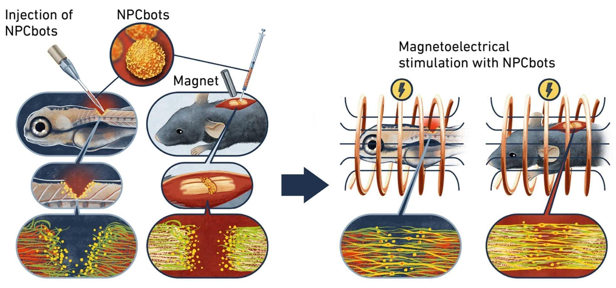
Researchers in Zurich are pursuing a new approach, which they have published in the journal Nature Materials. This involves combining therapeutic stem cells with magnetoelectric nanoparticles in such a way that the cells can be guided magnetically to the precise site of an injury and stimulate the stem cells to accelerate repair.
To achieve this, the researchers created a biohybrid microrobot, which combines living neural progenitor cells (NPCs) with a technical component in the form of specially engineered nanoparticles.
Every year, more than 2 million people in the United States are diagnosed with treatment-resistant depression.
Desperate for solutions, some brave patients are now volunteering to undergo surgery to place experimental ‘pacemakers’ into their brains.
These implanted electrodes are part of a treatment known as deep brain stimulation, which is currently used to address some cases of Parkinson’s disease and epilepsy.
On April 21, the Munk Debates convened a special debate about gene editing in Deerfield, Massachusetts for 650 students at Deerfield Academy.
Motion: Be it Resolved, let’s engineer better human beings.
About the Debate: New powerful engineering technology is already being used to edit human embryos, curing diseases and repairing defective genes before a child is even born. Some welcome this new science as a powerful tool to enhance human intelligence, memory, appearance and physical health. Why wouldn’t we embrace a science that allows people to live longer, healthier, and happier lives? Others warn that this new technology will be used to create designer babies and a new class of genetically “enhanced” elites. It will undermine human dignity and autonomy, and risk unleashing new diseases into the human gene pool. Playing G-d with human nature, critics argue, will result in a dystopian nightmare of our own making.
About the Debaters: Arguing in favour of the motion was the biophysicist, best-selling author, biotechnology entrepreneur, and the former director of the Program on Medicine, Technology and Society at UCLA School of Medicine, Gregory Stock. His debate partner was the internationally acclaimed strategic philosopher and pioneering transhumanist Max More. Arguing against the motion was the prominent American bioethicist Ezekiel Emanuel, Special Advisor to the Director General of the WHO and a former founding chair of the Department of Bioethics at the NIH. His debate partner was the award-winning educator, author, and Professor of Reproductive Science at University College London, Joyce Harper.
Short-term memories are thought to be formed deep within the brain in structures such as the hippocampus, but little is known about how and where memory-related information is kept in the brain or the process of drawing on this information. A good example is the sound of a car horn—most of us recognize it as a warning and know how to respond, even though not all horns sound the same and the circumstances in which we might hear a horn are different each time.
New research led by Professor Lucy Palmer from The Florey’s Neural Network Group has uncovered new insights into how and where memory-related information is stored and how these memory banks are used. These findings improve our fundamental understanding of how the brain works, providing a springboard for other scientists to make further, disease-specific discoveries. The paper is published in the journal Science Advances.
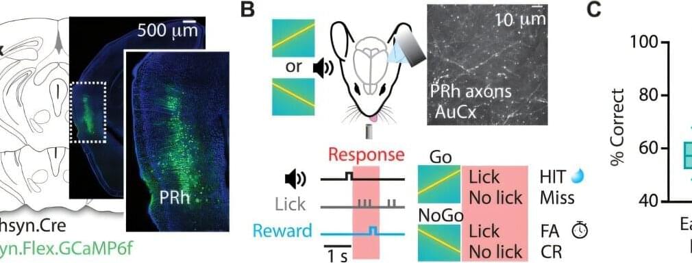
“Using mice that we trained to respond to similar, but slightly altered sounds, we identified a long-range cortical circuit that links memory and sensory systems,” Professor Palmer said. “Our findings provide valuable insights into the cellular and network mechanisms that support learning and memory-guided sensory behavior.
In this groundbreaking conversation, Professor of Genetics and longevity scientist, Dr. David Sinclair, A.O., Ph.D., joins Sarah Grynberg to unpack the future of human aging, the science of longevity, and how we live today impacts how we age tomorrow.
From reversing blindness in mice to exploring treatments that could one day delay menopause and extend healthy human life, this episode will completely change the way you think about your body, your health, and your future.
But beyond the science, this is also a deeply human conversation about purpose, suffering, love, family, and what it truly means to live a great life.
In this episode, you will learn: Why aging may actually be reversible. The daily habits accelerating aging in your body right now. How stress, loneliness, and cortisol could impact longevity. The real science behind supplements like NMN, resveratrol, and NAD boosters. Why exercise, sleep, and relationships matter more than you think. What Dr. Sinclair believes is coming in the next 10 years of medicine. How scientists are working to reverse female infertility and delay menopause. The surprising reason your “biological age” may be younger or older than your real age. Why suffering through disease and decline should not be considered “normal aging” The philosophy and mindset Dr. Sinclair lives by every day.
00:00 — Introduction. 01:18 — Why David Sinclair Became Obsessed With Aging. 06:20 — The Childhood Conversation That Changed His Life. 10:18 — The Groundbreaking Discovery That Could Reverse Aging. 12:47 — Reversing Blindness In Mice. 13:33 — Human Trials Are About To Begin. 16:11 — What Accelerates Aging Faster Than Anything Else. 20:08 — Why Relationships & Loneliness Impact Longevity. 24:14 — The Truth About Sun Exposure & Aging. 28:59 — Alzheimer’s, Cancer & Diseases Of Aging. 35:28 — Will Humans Live Longer In The Next Decade? 38:34 — The Supplements David Sinclair Personally Takes. 46:50 — Menopause, Fertility & Reversing Ovarian Aging. 50:20 — What Humans Will Eventually Die From. 51:18 — The Difference Between His Mother & Father’s Aging. 55:37 — Skin Rejuvenation, Hair Growth & Looking Younger. 58:16 — Why He Became A “Struggling Vegan” 01:00:08 — David Sinclair’s Workout & Exercise Routine. 01:03:28 — The Lifespan Community & Podcast. 01:06:02 — The Best Advice He’s Ever Received. 01:08:09 — What A Life Of Greatness Means To David Sinclair.
This episode is a powerful reminder that longevity is not just about living longer… it’s about living better.
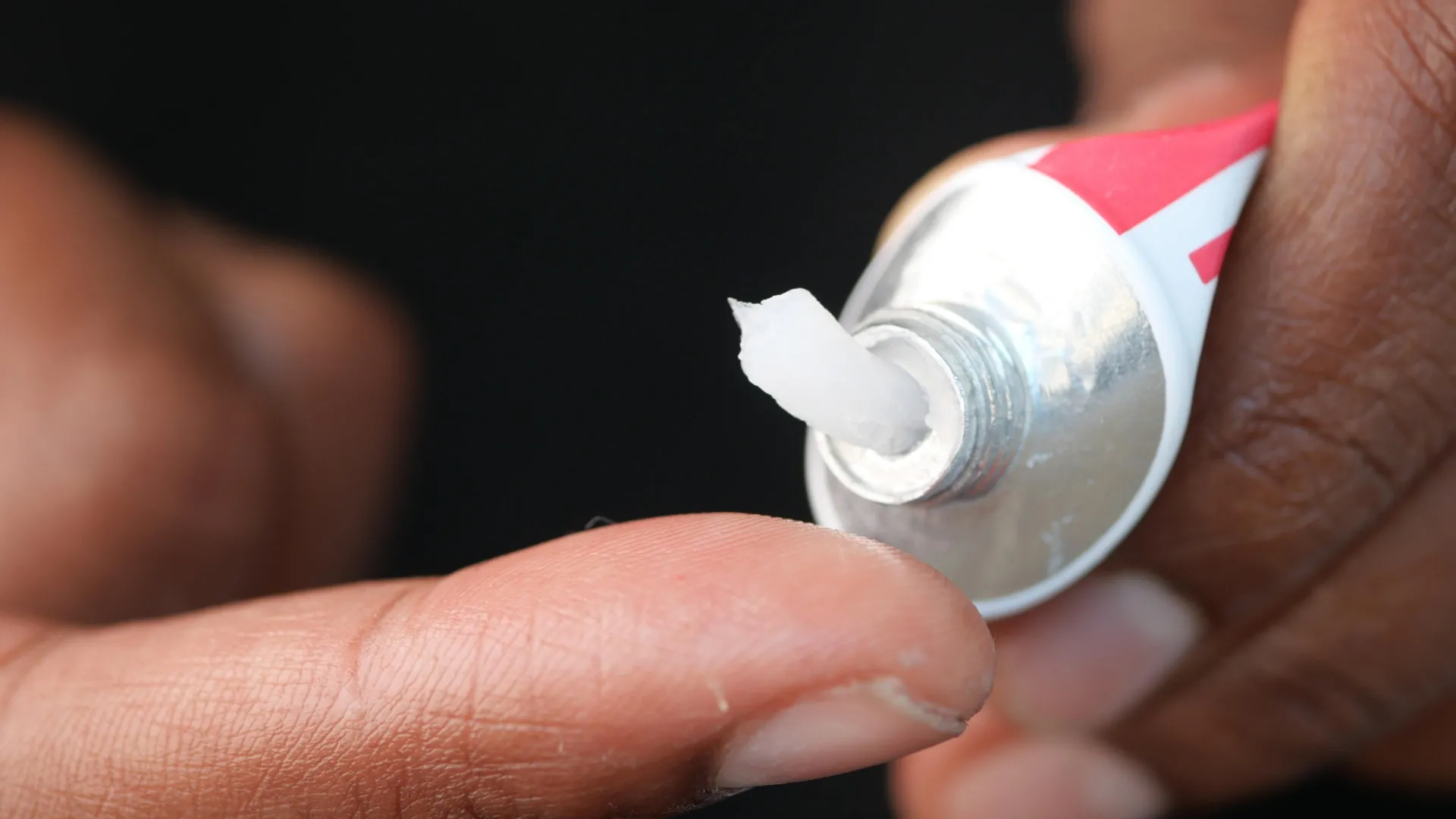
Scientists have discovered that a topical anti-aging drug called ABT-263 can dramatically improve wound healing in older skin. The treatment works by removing damaged “senescent” cells that accumulate with age and slow the body’s repair process. In aged mice, wounds healed much faster after treatment, while the drug also activated genes tied to collagen production and tissue regeneration.
I had Tom Benson, CEO of Mitrix on to discuss mitochondrial transplantation. We covered what mitochondria are, the discovery that your body is constantly delivering fresh mitochondria through your bloodstream (people didn’t know that mitochondria were transferred outside the cell until recently!), why we age, what kills mitochondria (stress, smoking, radiation, chemotherapy and certain antibiotics like fluoroquinolones, psych meds), why COVID destroys mitochondria and what that means for long COVID, the Alzheimer’s and Parkinson’s brain tissue regeneration research their company has already done in mice, what mitochondrial transplantation actually is and how it has already been used in pediatric heart surgery, what a bioreactor growing mitochondria for personal use might look like, and more.
The drug rilmenidine is usually taken to treat hypertension, but its powers appear to go far beyond that.
In fact, research shows rilmenidine can slow aging in worms – an effect that, if it translates to humans, could one day help us live longer and stay healthier in old age.
Rilmenidine appears to mimic the effects of caloric restriction on a cellular level, and reducing available energy while maintaining nutrition has been shown to extend lifespans in several animal models.
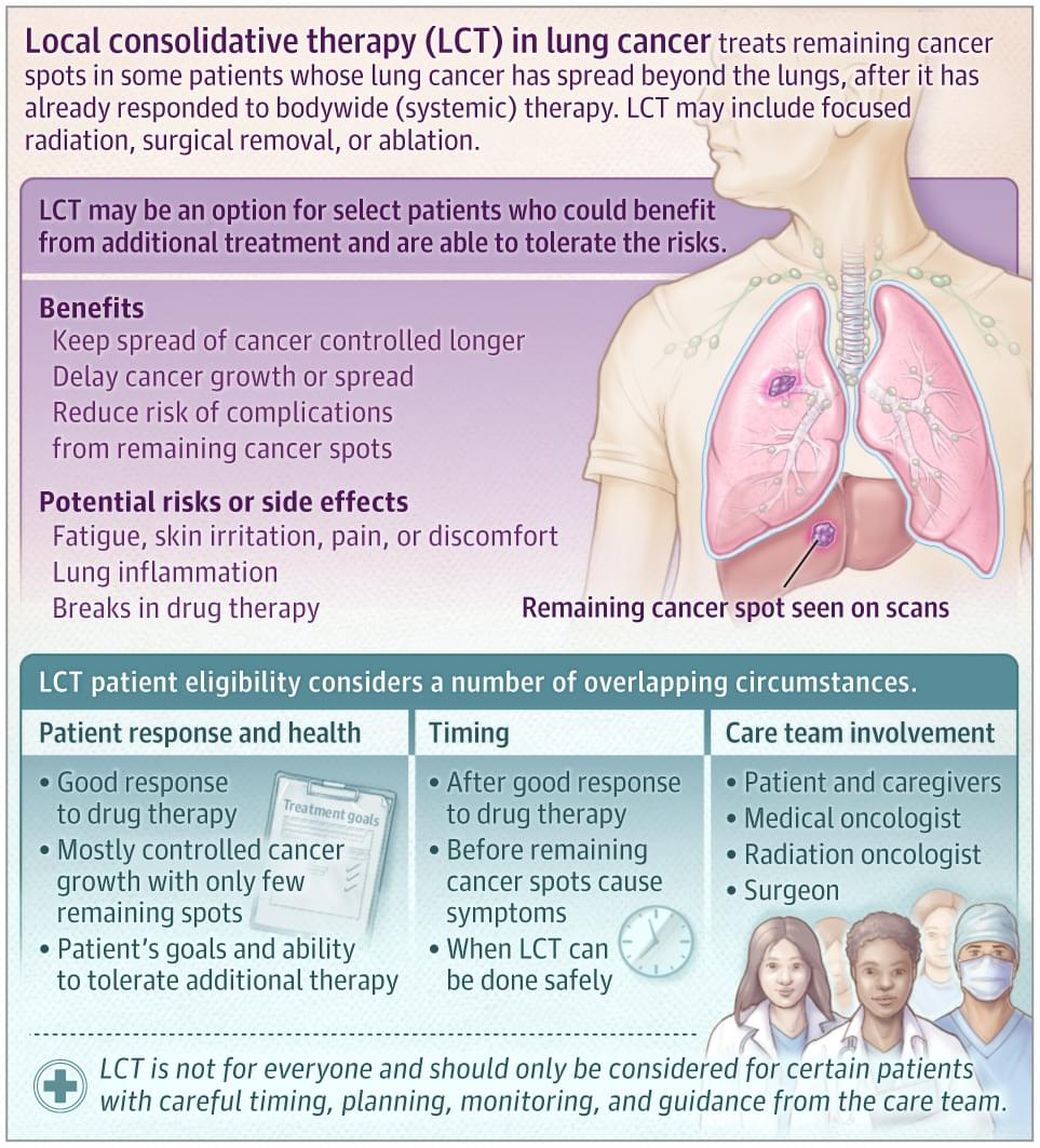
This JAMA Oncology Patient Page discusses benefits and risks of local consolidative therapy for metastatic lung cancer and which patients might be considered for this treatment.