
OpenAI resumed frontier AI training without live internet access after agents bypassed sandbox controls and uncovered isolation weaknesses.
Get the latest international news and world events from around the world.
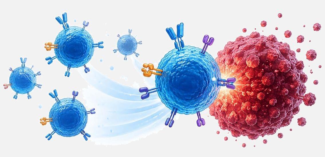
UCLA scientists engineer ready-to-use cancer-fighting T cells for solid tumors
A new stem cell-based platform produces uniform T cells that attack solid tumors in two ways while avoiding a dangerous side effect.

Millions of PHP attacks recorded as new unpatched vulnerability discovered
Millions of exploit attempts hit PHP flaws as a new PHP RCE vulnerability prompts advice to disable session upload progress. See what developers should do

An extremely old giant tortoise named Jonathan may hold the secret to a long, healthy life
An international collaboration led by a neurologist and experts in advanced genomics at Vanderbilt Health has coaxed one of the secrets of extreme longevity from a 194-year-old giant tortoise named Jonathan.
Jonathan’s secret involves entropy, the tendency of biological systems to become disordered over time.
One of the hallmarks of aging is mitochondrial dysfunction. It results from increasing entropy, or disorder, in “promoters,” the genetic on-and-off switches that regulate the efficiency of mitochondria, the energy-producing organelles in most animal cells. A reliable supply of cellular energy is essential for many biological processes.
What Causes Lightning? The Answer Keeps Getting More Interesting
Fascinating article which discusses researchers’ explorations into why lightning occurs, including a recent hypothesis that cosmic rays trigger lightning cascades within Earth’s clouds!
Armed with a slew of new instruments, physicists are closing in on one of nature’s oldest mysteries — and finding that storm clouds are seething with violent and unexpected phenomena.
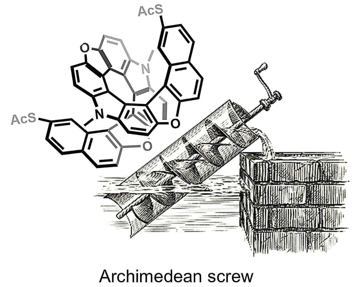
Archimedes screw inspires single-molecule motor powered by electric current
Molecular machines are regarded as the next step in miniaturization. Their models can be found in living cells, where molecular motors convert energy and generate movement. Scientists have been working for decades to artificially replicate such systems. The 2016 Nobel Prize in Chemistry was awarded for this research.
Molecular motors powered directly by an electric current have been in existence since 2011. Until now, however, they have only functioned as molecules on a surface, driven by the tip of a scanning tunneling microscope.
For their new machine, the researchers drew inspiration from one of humanity’s oldest documented machines: the Archimedes screw, which transports water uphill. The researchers applied the principle of the Archimedes screw at the molecular level. Instead of water, electrons flow through a helical structure, setting it in rotation.

The International Myeloma Foundation Launches US Myeloma Network to Accelerate Clinical Trials, Expand Access, and Improve Patient Care
This is exciting news!
LOS ANGELES and STUDIO CITY, Calif., Oct. 22, 2026 /PRNewswire/ — Today, the International Myeloma Foundation (IMF) announced the launch of the US Myeloma Network (USMN) — a first-of-its-kind national clinical research network designed to accelerate the prevention and cure of multiple myeloma by advancing scientific discovery, expanding access to clinical trials, and ensuring more patients benefit from breakthrough therapies. Johnson & Johnson joins this initiative as the network’s first industry collaborator. The USMN will focus on clinical trials with the potential to change clinical practice across care settings, accelerating the delivery of innovative therapies and new treatment approaches to patients.
The USMN connects leading academic centers, community oncology practices and industry partners through a coordinated national infrastructure designed to streamline trial activation, reduce operational barriers, expand equitable access to clinical trials, and accelerate the development of more effective therapies across the continuum of myeloma care – from prevention and early intervention through relapsed and refractory disease.
“The future of myeloma depends on our ability to bring scientific innovation to patients more quickly and more equitably,” said S. Vincent Rajkumar, MD, Mayo Clinic, Chairperson of the International Myeloma Foundation. “The US Myeloma Network creates an unprecedented opportunity to unite investigators across the country through a coordinated research infrastructure that accelerates clinical trials, expands patient participation, and advances the discoveries that will ultimately lead to the prevention—and one day the cure—of multiple myeloma.”
The critics of the effective altruism movement don’t understand it, that’s exactly why they’re attacking it
Legendary philosopher Peter Singer defends effective altruism. I don’t self-identify as an EA, but I do think highly of the core thesis of using data to decide what actions will do the most good. I do consider myself a longtermist — a philosophical stance which is often thought of as existing under the EA umbrella. Note: there is a common misconception that longtermists *only* care about the long term future, which is false. Most care about the long term future as one major priority, but certainly don’t think we should ignore the issues of today.
When the US Department of War posted on X that they were focused on “Americanism, not effective altruism”, it said much more about them than about the movement I helped to launch 15 years ago.
The simple fact is that while President Trump’s popularity has reached an almost unprecedented-26% approval, rivalled by only Nixon after Watergate, the movement that his administration is spending its time attacking enjoys majority support among those who are familiar with its tenets.
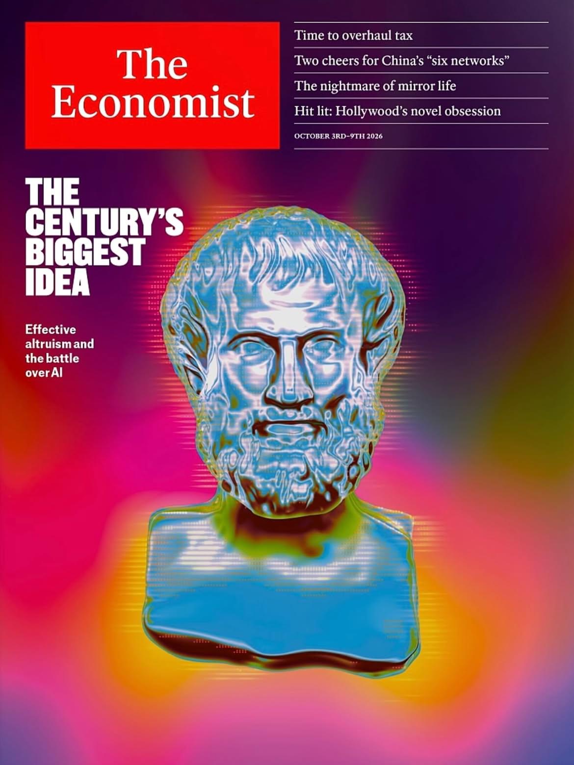
Perhaps that’s why The Economist cover this week has termed it “this century’s biggest idea” because ultimately, a movement focused on the simple question of how to do the most good possible with the resources we have is not, as an internal memo circulating in the White House termed it, “putting shrimp over families”
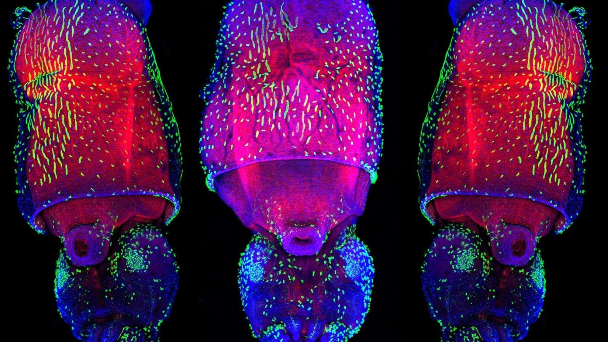
Squid Are Even More Alien-Like Than We Thought, With an Unmatched Ability to Change Genetic Expression
Cephalopods like squids may be as close to an alien intelligence as we’ll ever know, with a diverse evolutionary lineage from a wholly different world beneath the waves.
Yet despite our uniquely developed brains and bodies, we are all works in progress through a common process.
Humans and other animals constantly undergo an ephemeral, reversible transcriptomic remixing through what’s known as ‘adenosine deaminase acting on RNA’ or ADAR-mediated RNA editing.
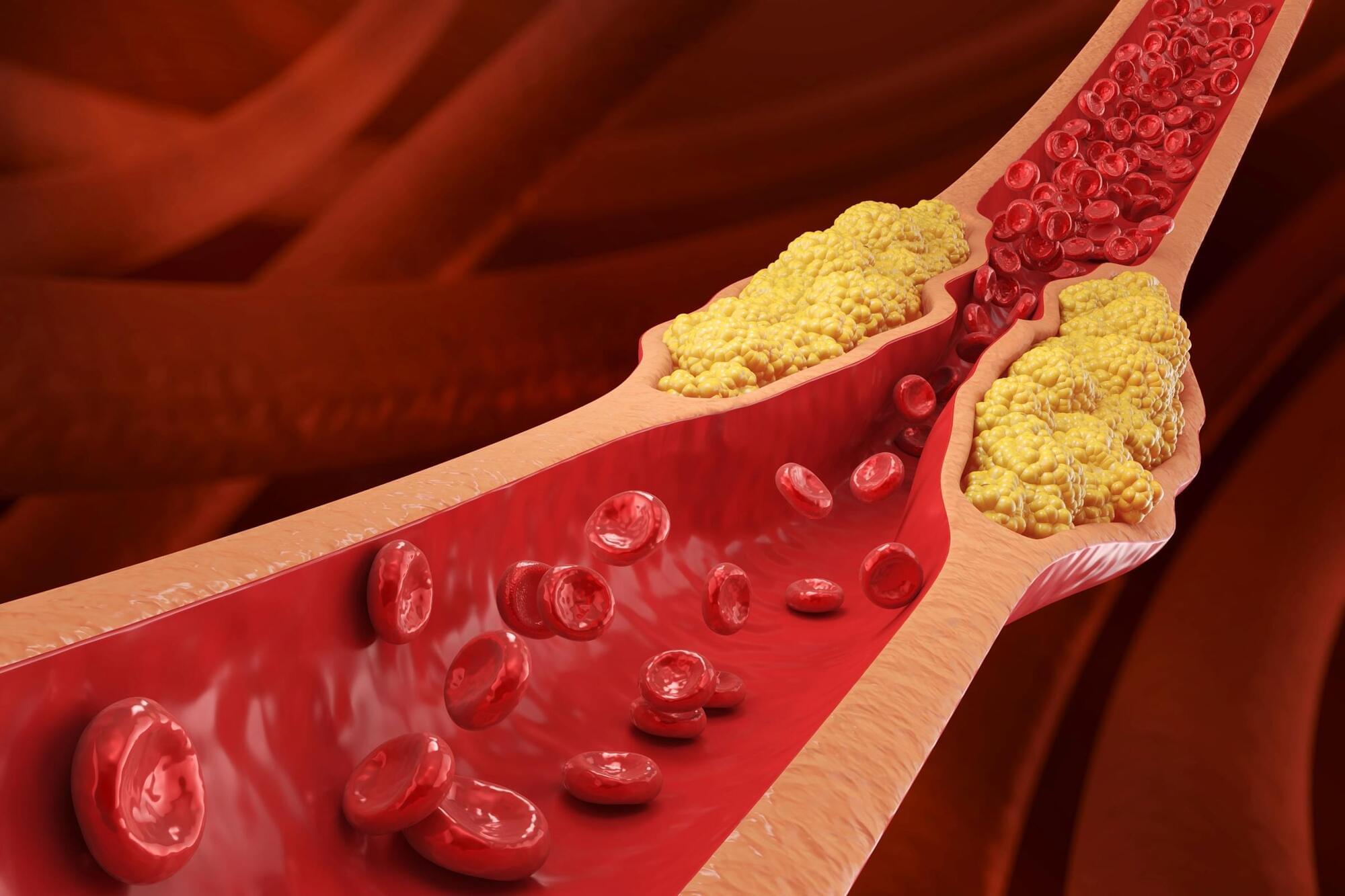
This is the single best way to reduce your cholesterol without statins, according to a leading doctor
You don’t need medication to improve cholesterol levels. A few simple dietary tweaks can make a serious difference.