Human hibernation seems like pure science fiction, but it could make interstellar travel a reality.

Scientists who use imaging to understand the brain’s complexity often focus on the strongest signals and ignore the rest. But this strategy, researchers warn, may reveal only the tip of the iceberg. A study published in Nature Human Behavior reveals that connections routinely overlooked as “noise” during neuroimaging data analysis can predict behavior with remarkable accuracy—and implicate entirely different brain networks. The finding could open many new targets for treating psychiatric illness, the researchers say.
“Many studies that rely on techniques like feature selection—which simplifies the brain down to a narrow slice—might only uncover a small part of the true neurobiology that underlies a given behavior,” says lead author Brendan Adkinson, Ph.D., an MD-Ph. D. student at Yale School of Medicine.
“Our study suggests that there may be multiple, non-overlapping networks capable of predicting a given behavior just as well.”

God is said to be all-powerful, all-knowing, all-good. But what is God’s private mental life like? Can we reach in to appreciate God as a supreme being? It may seem absurd, or arrogant, for finite human beings to strive to imagine what an infinite God is like and even what God may feel like privately and inside. But that is what we do.
John Charlton Polkinghorne KBE FRS was an English theoretical physicist, theologian, and Anglican priest.
Watch the Big Questions About God playlist:
• Big Questions About God — Closer To Truth…
https://closertotruth.podbean.com.
Get access to over 5,000 videos by signing up for a free Closer To Truth membership:

Intellectually engaging and stimulating activities like reading, writing, and learning new languages are linked to a lower risk of Alzheimer’s disease and mild cognitive impairment in later life. The corresponding study was published in Neurology.
“Our study looked at cognitive enrichment from childhood to later life, focusing on activities and resources that stimulate the mind. Our findings suggest that cognitive health in later life is strongly influenced by lifelong exposure to intellectually stimulating environments,” said study author, Andrea Zammit, PhD, of Rush University Medical Center in Chicago, in a press release.
For the study, the researchers analyzed data from 1939 adults with an average age of 80 years old who were dementia-free at the start of the study. They were followed for around eight years.
In this video, leading philosophers and neuroscientists defend the view that the mind purely physical?
Starring some of the very experts who anti physicist quote such as Bob Kirk (Zombie argument) and Frank Jackson (Marys room argument) who have now turned to physicalism, as well as the most cited neuroscientists in the world, Karl Friston and other leading scholars such as Ned Block, David Papineau, Richard Brown, Ken Williford, Anil Seth and Marc Solms, we examine the strongest case for physicalism—the view that everything about the mind can ultimately be explained in terms of the physical brain.
We take on some of the most famous anti-physicalist arguments, including: The Hard Problem of Consciousness, Knowledge arguments (e.g., Mary’s Room), Philosophical zombies Dualist intuitions about the self and panpsychism.
Do these arguments really show that consciousness is non-physical—or do they rely on misconceptions about how the brain works?
This video breaks down complex ideas into clear, rigorous explanations while challenging some of the most popular objections to physicalism.
If you’re interested in philosophy of mind, consciousness, neuroscience, or the nature of reality itself, this is for you.
00:00 Introduction

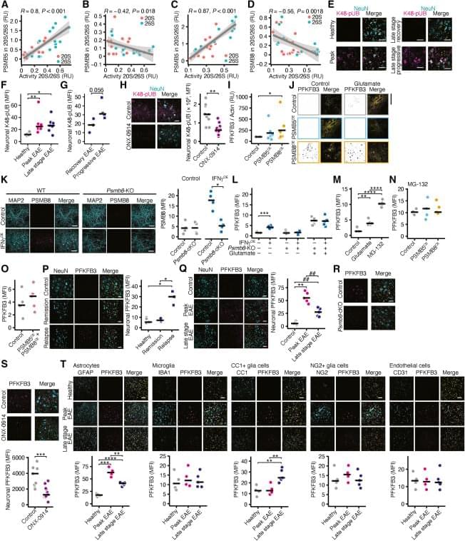
Now online! The immunoproteasome disturbs neuronal metabolism and drives neurodegeneration in multiple sclerosis: (Cell 188, 4567–4585.e1–e12; August 21, 2025)
Now online! (Cell 188, 4567–4585.e1–e12; August 21, 2025)
During post-publication review of our article, we, the authors, identified several errors in figure assembly and annotation affecting representative images and sample size reporting. These issues are limited to figure presentation and do not affect the underlying data, quantification, or conclusions of the study.
In Figure 2G, incorrect representative images were inadvertently used for the interferon-γ-OE and PSMB8-OE glutamate conditions. The correct images have now been inserted.