Toggle light / dark theme

Plasticity as a therapeutic target for improving cognition and behavior in Down syndrome

Early intervention and environmental optimization have been central to management of Down syndrome (DS) and much of current treatment is still focused in strategies that involve early education plans. This approach has provided significant improvements for Down syndrome but it is not providing a full success. The discovery of an increasing number of genes and molecular pathways linked to intellectual disability and involving a range of synaptic and plasticity-related mechanisms has open new treatment opportunities that focus on targeted treatments boosting neural plasticity. We here discuss some of these approaches, focusing on the effects of environmental enrichment and on the discovery of pharmacological therapies showing beneficial effects even in some clinical trials in adult individuals with Down syndrome. Targeting plasticity impairments in DS is thus a promising strategy to promote cellular mechanisms involved in learning and memory within key cognitive brain region and could lead to improved connectivity.

Keywords: EGCG; Environ-mimetic drugs; Environmental enrichments; Epigenetics; Neuronal plasticity.

© 2020 Elsevier B.V. All rights reserved.

Brain Delivery of Antibody-Derived Biologicals for Alzheimer’s Disease: An Updated Narrative Review

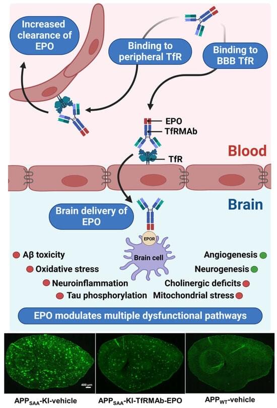
Antibodies directed against β-amyloid (Aβ) have been developed for the treatment of Alzheimer’s disease (AD). However, the in vivo central efficacy is reduced by the poor penetration of antibodies across the blood–brain barrier (BBB). In addition, these antibodies have been associated with adverse effects like amyloid-related imaging abnormalities. Thus, the development of new antibody-based therapies for AD with improved transport across the BBB may improve efficacy and reduce adverse effects. Antibodies targeting the BBB transferrin receptor (TfR) are able to cross the BBB through receptor-mediated transcytosis, producing a global distribution throughout the brain.

Astrocytes can gain neural stem cell properties after brain injury

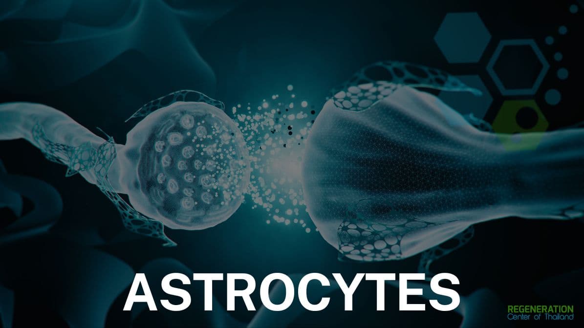
New research shows that specific types of brain cells become active after brain injuries and exhibit properties similar to those of neural stem cells. Astrocyte plasticity might correlate with the upregulation of the Galectin 3 protein, which may significantly contribute to discovery of additional biomarkers. The study discovered that a specific protein regulates these cells and could be a target for therapy and contribute to development of better treatments options for brain injuries. The loss of neurons, which subsequently causes impairment of brain function, is caused by the onset and progression of neurological disorders, like strokes, spinal cord injuries and neurodegenerative diseases such as Parkinson’s, Alzheimers / Dementia, ALS and MND. Effective treatment options still need to be improved. However, preclinical research has shown a promising response involving reactive astrocytes, a specific type of glial cell, which is a crucial part of the nervous system alongside neurons. Microglia and Glial cells are regarded as a safeguard for neurons, demonstrating the ability to resume cell proliferation, a mechanism essential for protecting the injury-affected brain from invasion by immune cells.[1]

Differentiation of Mesenchymal Stem Cells to Neuroglia.

Given the importance of astrocyte proliferation, these findings are relevant for understanding how changes in cerebrospinal fluid composition (upregulation of Galectin 3 protein) support the maintenance of astrocyte plasticity in the brain. Identifying Galectin 3 protein as an inducer of astrocyte plasticity has helped discover other biomarkers that offer beneficial modulation inside the injured brain parenchyma. These regulators of astrocyte proliferation after acute injury offer great promise for the future clinical applications of these biomarkers as indicators for detecting a beneficial reaction of glial stem cell therapy or help identify the presence of other cells with stemness potential in an injured patient’s brain [5].

Radiologic analysis of large vestibular schwannoma position on surgical outcomes

Large vestibular schwannomas (VS) often compress the brainstem and differ in their relation to the internal auditory canal (IAC); the significance of these radiographic features on postoperative outcomes remains unclear. This study quantifies the impact of brainstem compression (BSC) and position relative to the IAC on surgical outcomes in VS.

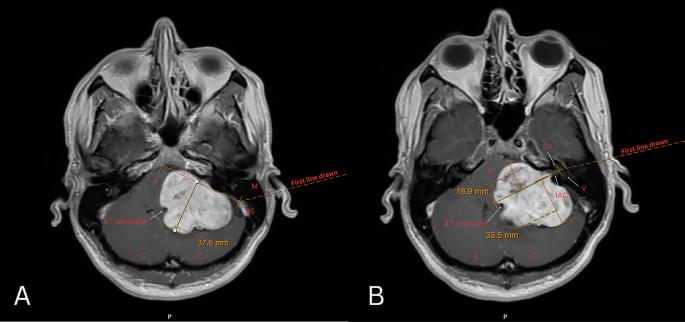
We retrospectively identified 116 patients with sporadic unilateral VS ≥ 3 centimeters (2017–2022). Neurofibromatosis 2 cases were excluded. BSC was quantified with MRI T1 post-contrast axial images as the perpendicular distance from the brainstem-cerebellum to the point of maximal compression. Anterior and posterior IAC extension were measured relative to a line bisecting the IAC from the porus to fundus. Outcomes included postoperative facial nerve (FN) function, extent of resection (EOR), and length of stay (LOS).

Greater anterior extension was associated with decreased EOR in univariate analysis (OR = 1.12, p = 0.03), but not after controlling for tumor size and age (OR = 1.09, p = 0.158). Greater BSC was associated with worse FN function at 2–3 weeks postoperatively on univariate (OR = 1.08, p = 0.036) and approached significance on multivariate analysis (OR = 1.07, p = 0.08). Posterior extension was associated with increased LOS in univariate (β = 217.57 min, p = 0.024), but not multivariate analysis. Neither anterior extension nor BSC were associated with LOS. Older age correlated with a lower rate of GTR and longer LOS in multivariate analysis (EOR: OR = 1.05, p = 0.003; LOS: β = 79.84 min, p = 0.026).

Your Brain Has Two Minds | NOVA | PBS

Neuroscientists are uncovering how communication between different parts of the brain creates the experience of being “you.”

📺 Watch the full episode here: https://www.pbs.org/wgbh/nova/video/y…

💙 Help us bring the universe closer to home. Your donation ensures that NOVA’s deep-space discoveries and quantum physics explorations remain free and inspiring for the next generation of scientists. Support fact-based storytelling today. https://bit.ly/48UnsrD

📰 Stay up to date on the latest science discoveries, full episodes, articles, videos, and more by signing up for NOVA’s newsletter here: https://to.pbs.org/4brCC7Y

▶️ Stream NOVA documentaries \& more with PBS Documentaries on Prime Video: https://amzn.to/4hXBlsu.

🛍️ Shop official NOVA 50th anniversary merch: https://nova-shop.org/

Brain Morphology Mediators of the Association of Childhood Trauma With Bipolar Disorder: An International ENIGMA Bipolar Disorder Working Group Study

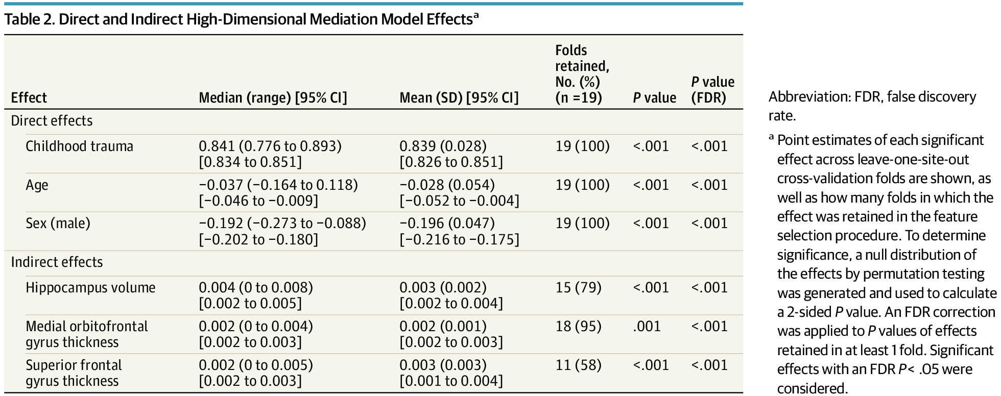
This case-control study analyzes the mediating effects of brain morphology on the association of childhood trauma with bipolar disorder.

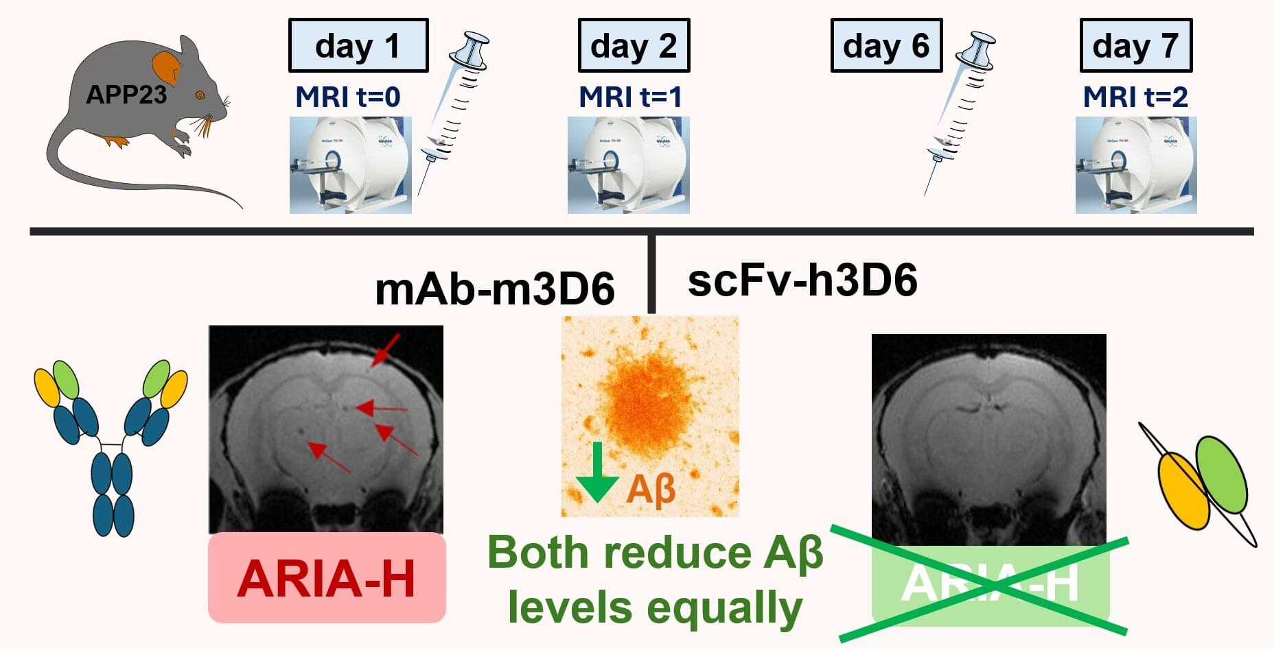
Antibody fragment prevents hemorrhages associated with new Alzheimer’s treatments

In 2025, the European Medicines Agency approved two antibodies for Alzheimer’s disease: lecanemab (LeqembiTM, from Biogen) and donanemab (Kisunla, from Eli Lilly and Co.), both based on immunotherapy (the use of molecules from the immune system to treat diseases). These antibodies, obtained in the laboratory, act against the Aβ peptide, a protein fragment that accumulates in the brains of patients with Alzheimer’s disease. Elimination of this protein by the immune system helps slow the characteristic cognitive decline of the disease.

These two antibodies are the first disease-modifying therapies for Alzheimer’s. They stop and, in some cases, even partially reverse this devastating condition. However, a frequent and characteristic side effect of these drugs is cerebral bleeding, detectable by magnetic resonance imaging. The brain does not have the molecules and cells that make up the systemic immune system, so the entry of antibodies into the brain is not desirable under healthy conditions, although it is necessary for these treatments to be effective.

The incidence of bleeding in clinical trials ranged from 10% to 27% of treated patients, with a particularly high incidence in individuals carrying a specific apolipoprotein allele: APOEε4. In Europe, these treatments can be administered only to people with one or no copy of the APOEε4 allele, a genetic variant associated with a higher risk of Alzheimer’s.

Long-range white-matter pathways enable efficient spontaneous neural activity propagation in the human brain

Efficient brain-wide communication requires neural activity to traverse long anatomical distances rapidly. Here we examine how propagation timing is jointly associated with spatial geometry, functional network organization, and long-range white-matter pathways and their microstructural properties. And we ask whether the same rules govern epileptiform and physiological activity. Using stereo-EEG and diffusion spectrum imaging from 47 epilepsy patients (26 males and 21 females), we quantified inter-regional propagation with two complementary delay estimators: event-based interictal epileptiform discharge (IED) traveling waves and continuous lagged-correlation delays during IED-free periods. We found that IED propagation traversing gray and white matter formed reproducible spatiotemporal motifs that deviated from randomized null models, indicating structured routing rather than random spread. Epileptiform and physiological propagation delays increased over short ranges but saturated at longer distances, indicating that geometry alone cannot account for long-range fast propagation. Beyond geometry, stronger structural connectivity and higher functional connectivity were associated with shorter delays, and intrinsic functional modules facilitated efficient communication: within-network propagation was faster than between-network propagation. Crucially, diffusion-derived quantitative anisotropy (QA) revealed a microstructural mechanism for long-range fast propagation: long-range white-matter tracts showed higher QA, and QA was positively associated with apparent propagation velocity. Together, these results identify convergent, architecture-dependent constraints on propagation timing that generalize across epileptiform and normal activity, providing a principled bridge between macroscale connectome organization and fast intracranial spatiotemporal dynamics.

Significance statement Efficient communication across long anatomical distances is fundamental for the human brain. By integrating stereo-EEG with diffusion spectrum imaging, this study shows that brain-wide information propagation is not determined by distance alone, but is critically supported by long-range white-matter pathways, their microstructural properties, and intrinsic functional network organization. We also find that both pathological epileptiform discharges and physiological spontaneous activity follow shared propagation rules, exhibiting distance saturation, structural facilitation, and preferential within-network transmission. These findings provide a microstructure-grounded account of how the human brain achieves fast, efficient large-scale communication, bridging macroscale connectome architecture with millisecond-scale neural dynamics.

/* */