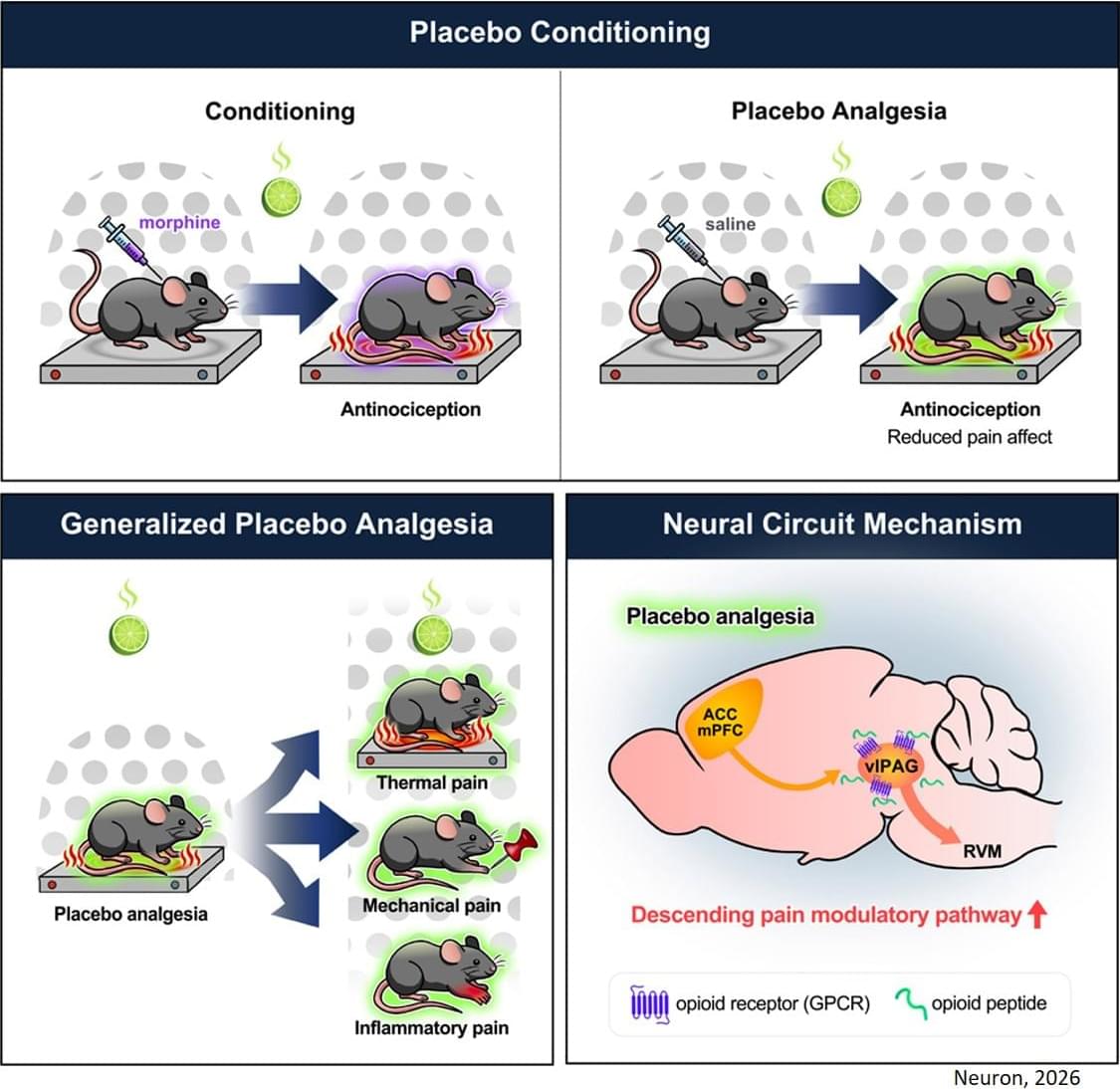
Toggle light / dark theme

New sensor could allow MRIs to see molecular-level changes

You’ve seen people sliding into the tube of a magnetic resonance imaging (MRI) machine on your favorite medical drama, or maybe you’ve been inside one yourself, waiting as the noisy scanner makes images of your brain, heart, bones, or other structures, which doctors use to identify injury or disease.

Since the 1970s, MRIs have been important diagnostic tools, combining a magnetic field and radio waves to produce snapshots of the body’s interior without using ionizing radiation, which can create health risks at higher doses. An MRI can typically capture changes in anatomy, but the molecular-level changes that could further aid understanding of disease have been beyond its reach.

Now, in a new article in Science Advances, University of California, Santa Barbara researchers report the invention of a modular, genetically encoded, protein-based sensor that enables MRI machines to visualize molecular activity inside cells—a development that could transform how scientists study cancer, neurodegeneration, and inflammation.

The neuroscience of hypocrisy points to a communication breakdown in the brain

Half of the participants received actual stimulation aimed at the ventromedial prefrontal cortex. The other half received a fake version of the treatment, known as a sham stimulation. After the procedure, all participants completed the same card game and judgment exercises.

The people who received the real brain stimulation showed a wider gap between their behavior and their judgments. By disrupting the normal function of the brain region, the researchers successfully made people more hypocritical. This proved that the ventromedial prefrontal cortex directly controls moral consistency.

These results suggest that moral consistency is not an automatic trait. It is a biological process that relies on the brain’s ability to sync up different types of information. “Our findings suggest that we should treat moral consistency like a skill that can be strengthened through deliberate decision making,” says senior author Hongwen Song of the University of Science and Technology of China.

Chemokines CXCL9 and CCL2 in Relation to Cerebral White Matter Disease, Cognitive Decline, and DementiaThe Northern Manhattan Study

This large cohort study showed that higher serum CXCL9 was associated with greater burden of white matter disease in the brain, independent of vascular risk factors, renal function, and genetic predisposition, supporting a role for CXCL9 in white matter pathogenesis.


Background and Objectives.

Prevalence of early-stage type 1 diabetes in young adults: a population-based cohort study

Individual variability in synaptic gene expression and synapse density in induced pluripotent stem cell–derived neurons predicted macro-scale alterations in gray matter volume and gamma-band activity in patients with Schizophrenia.

SIRS2026.


This genetic association study tests whether genetically driven variability in excitatory neurons’ transcriptome and synapse density in patient-derived neurons in vitro explain individual changes in cortical morphology, electrophysiology, and cognitive impairments in vivo.

Development of a Diagnostic Autoantibody Assay to a Consensus Motif for the Risk Prediction of Epstein-Barr Virus–Related Multiple Sclerosis

Background and ObjectivesMultiple sclerosis (MS) is a chronic progressive, demyelinating autoimmune CNS disease. Autoantibodies to the motif P-(SA)-x-(SGA)-R-(SN)-(LRKH) are a class of predictive markers specific to MS that could add to emerging…

Genetically modified marmosets as a model for human deafness provide a foundation for future gene therapies

Why are some people unable to hear from birth, even though their inner ear appears intact? One possible cause lies in the so-called OTOF gene. It plays a central role in transmitting sound signals from the hair cells to the auditory nerve. Without this function, acoustic information does not reach the brain.

Researchers from the German Primate Center—Leibniz Institute for Primate Research, the University Medical Center Göttingen, and the Max Planck Institute for Multidisciplinary Sciences have now, for the first time, generated marmosets in which this gene has been knocked out precisely. The animals are healthy and develop normally, but are deaf from birth. This provides the first primate model that realistically replicates key characteristics of human deafness. The results are published in Nature Communications.

Hearing loss is one of the most common congenital sensory disorders in humans. A major cause is a defect in the OTOF gene. This gene ensures that the protein otoferlin is produced in the inner ear. This protein is necessary for sound signals to travel from the hair cells to the auditory nerve. Without it, the ear still functions externally, but the signals do not reach the brain.

Sean Carroll & Philip Goff Debate ‘Is Consciousness Fundamental?’

This debate took place in Marist College on Friday September 8th 2023. It was one of the public components of a conference on the topic of panpsychism organised by Andrei Buckareff and Philip Goff, as part of the Templeton funded project ‘Panpsychism and Pan(en)theism: Philosophy of Religion meets Philosophy of Mind.’ https://sites.google.com/view/panpsyc

Filmed and edited by Jay Shapiro.

How Hair Cells in the Ear Actively Respond to Sound

Tiny hair cells located in the inner ear help us hear and maintain balance. On top of each hair cell is a hair bundle, a sensory organelle that converts mechanical input from sound or movement into electrical output, which is then passed on to the brain. Previous research has shown that hair bundles aren’t simply passive entities. They actively oscillate to amplify weak audio signals or to tune into specific frequencies. Biologists have also observed bundles oscillating in the absence of stimuli. Models have tried to capture this bundle behavior, but the connection between active oscillation and the audio response has not been made clear. A new thermodynamic model of energy flow within hair bundles suggests that they work like tiny machines [1]. Depending on the stimulus, the bundles either extract power from incoming sound waves or inject power into them—corresponding, respectively, to sensing or amplifying a stimulus.

In the inner ear, an active process called cochlear amplification helps humans (and other mammals) hear the faintest of sounds. When a faint whisper enters the ear, for example, the outer rows of hair cells respond to the weak signal by moving in a way that amplifies the sound waves for the inner hair cells, which are the ones that send a message to the brain. Molecular motors propel the movement or twisting of hair bundles required for these functions.

Previous work has explored how much energy a hair cell consumes to drive bundle oscillations, but the resulting models have typically assumed that bundles are moving spontaneously—that is, in the absence of external stimuli. Roman Belousov from the European Molecular Biology Laboratory in Germany and his colleagues have developed a stochastic thermodynamic model that includes an energy input from sound waves. “Instead of just looking at how a hair bundle moves on its own, we wanted to add what happens when it interacts with sound,” Belousov says.

/* */