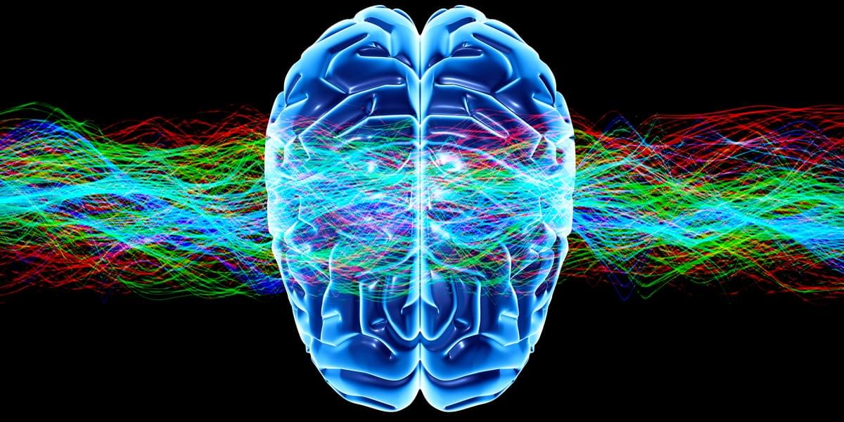
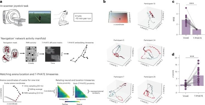
Busch et al. use nonlinear neural manifolds to help humans gain rapid control over a noninvasive brain–computer interface, allowing them to learn how to play a video game with real-time fMRI neurofeedback from cognitive brain regions.
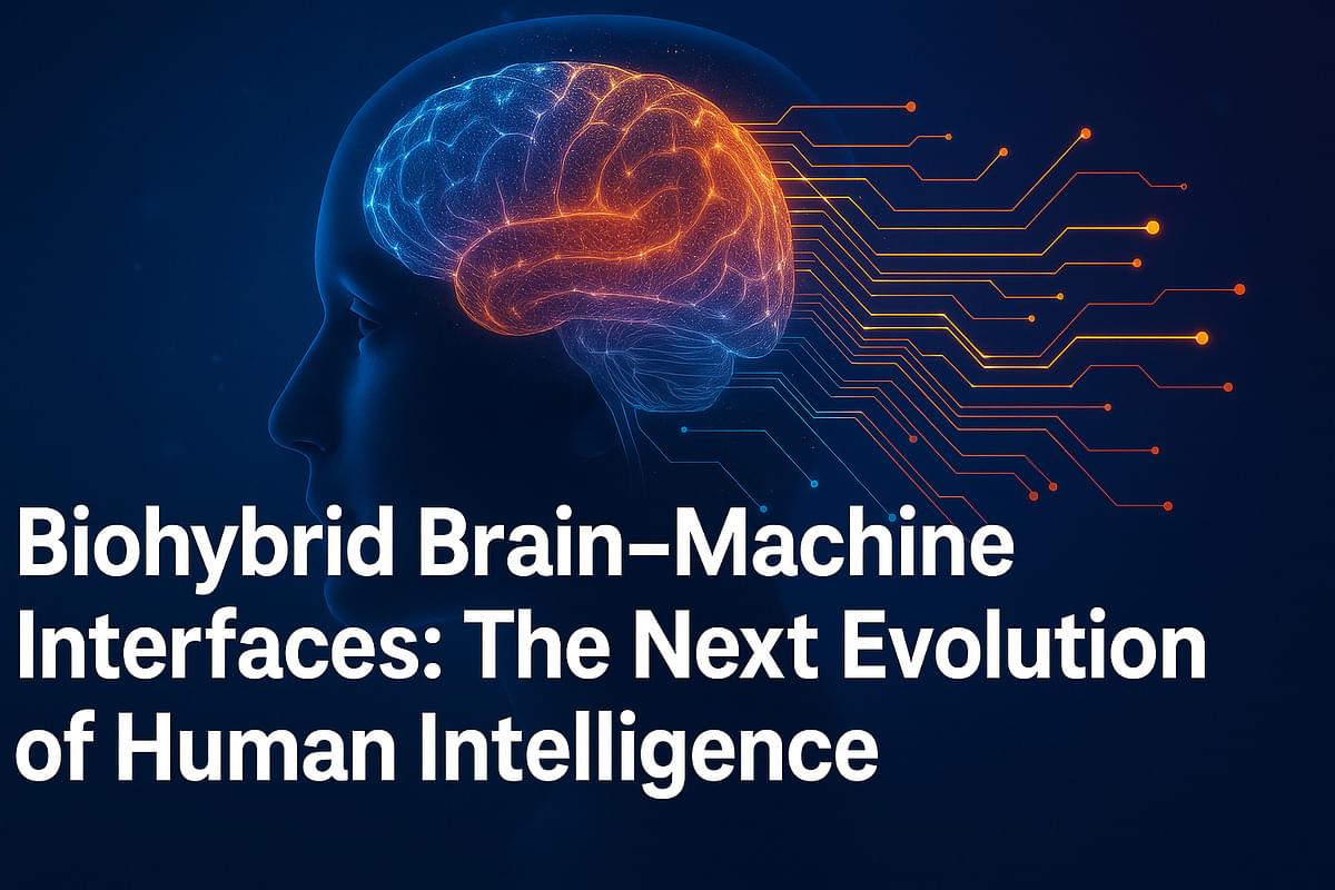
Brain–machine interfaces (BMIs) are no longer just science fiction; they are the gateway to a future where thought itself can interact directly with technology. These systems read the brain’s electrical activity and, in turn, stimulate neurons — forming a two-way communication link between biology and machines.
In just a few decades, BMIs have evolved from laboratory curiosities into one of the fastest-growing frontiers in science and engineering. The possibilities are staggering. In the future, neural interfaces could restore vision to the blind, enable paralyzed individuals to move again, facilitate seamless communication between human brains and artificial intelligence, and ultimately power virtual realities that are indistinguishable from the physical world.
This convergence of biology, computing, and neuroscience marks the dawn of a new era — one where the boundaries between human and machine begin to blur.
Millions of people worldwide are periodically or chronically affected by gut-related conditions, such as irritable bowel syndrome (IBS), gastroesophageal reflux disease (GERD) and gastroenteritis. Uncovering the physiological and biological processes that contribute to gut health could thus be highly valuable, as it might help devise more effective interventions to prevent and treat these ailments.
The transit of food, fluids and waste through the intestine is known to be coordinated by various interacting systems in the body, including gut wall muscles, neurons in the gastrointestinal tract and hormones. A growing body of research has also been exploring the crucial contribution of bacteria and other microorganisms residing in the digestive tract, which are collectively referred to as the gut microbiome.
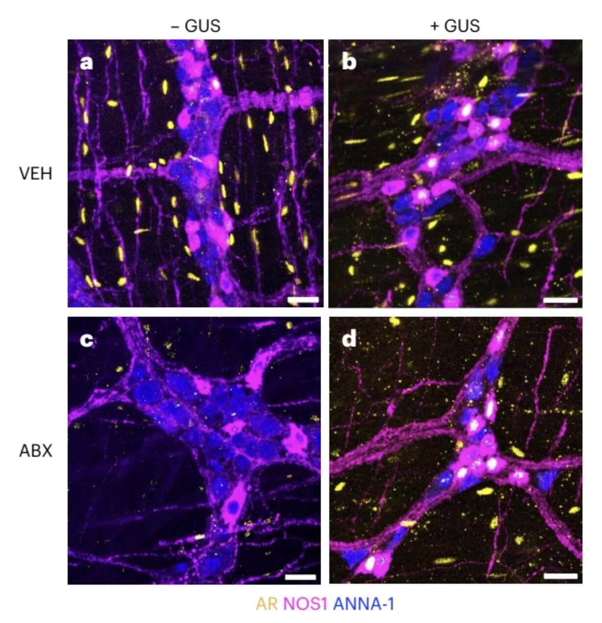
Researchers at Boston Children’s Hospital, Harvard Medical School, the University of North Carolina at Chapel Hill and Laval University recently carried out a study aimed at better understanding how these gut microbes interact with specific sex hormones and nerve cells that control the movement of muscles in the intestines.
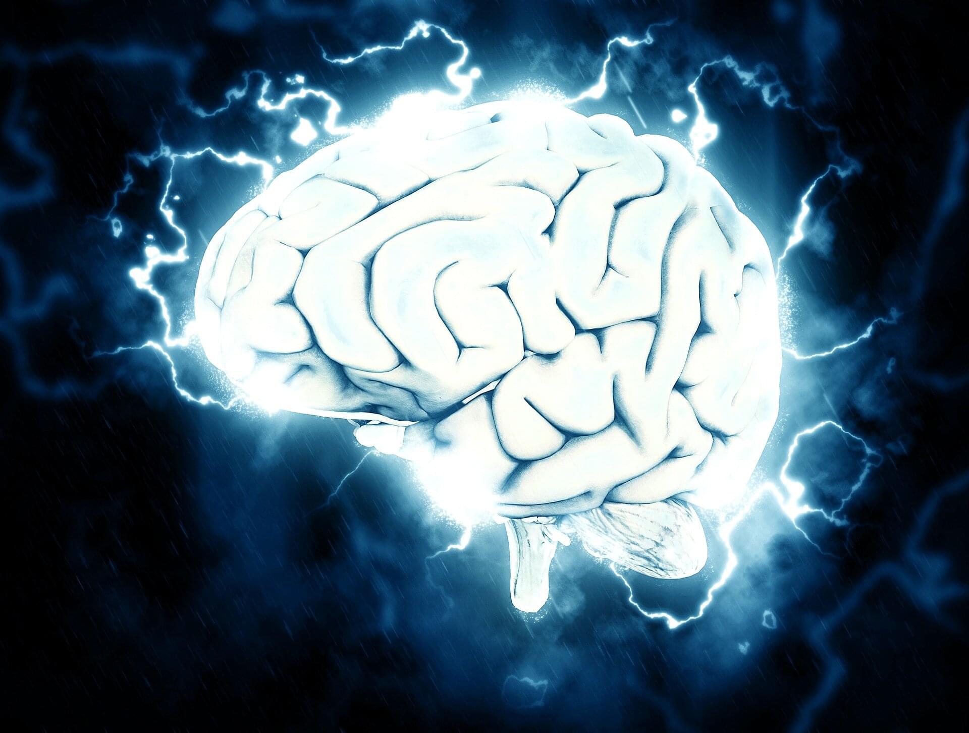
Researchers at the Icahn School of Medicine at Mount Sinai have identified distinctive patterns in how the brain transitions between activity states in people with depression, providing new insight into why depressive symptoms can feel persistent and difficult to overcome.
Published online in Nature Communications, the study combined advanced neuroimaging techniques with mathematical modeling to examine how the brain moves between functional activity states over time. The findings suggest that depression may involve a form of “brain-state entrapment,” in which the brain becomes more likely to enter certain patterns of activity and less likely to transition out of them.
“Many patients describe depression as feeling stuck in negative patterns of thought, mood and behavior,” said Yael Jacob, Ph.D., assistant professor of psychiatry at the Dennis S. Charney, MD, Depression and Anxiety Discovery Center at the Icahn School of Medicine at Mount Sinai and senior author of the paper. “Our findings suggest that this experience of being ‘stuck’ may reflect measurable changes in the brain’s underlying dynamics.”
To try everything Brilliant has to offer—free—for a full 30 days, visit https://brilliant.org/ArtemKirsanov/. You’ll also get 20% off an annual premium subscription.
===== My name is Artem, I’m a neuroscience PhD student at Harvard University. 🌎 Website and Social links: https://kirsanov.ai/ 📥 \.
To learn more, please visit the YouTube Help Center: https://www.youtube.com/help
Your brain is running on twenty watts right now. The power of a dim lightbulb. And yet it contains the entire eight-hundred-million-year history of life’s most improbable experiment — the experiment of intelligence itself. In this episode, we follow that experiment from its very beginning: from the first bacterium that navigated a chemical gradient in the ancient ocean, through the nerve nets of jellyfish, the distributed arms of the octopus, the tool-making crow, the grieving elephant, the dreaming mammalian brain — all the way to the only creature that has ever turned its intelligence on the question of where intelligence came from. This is not a story about the human brain. It is a story about what matter does when evolution pressures it long enough and hard enough. It is the deepest origin story you have.
/ @theevolutionoflife2026 Subscribe to the channel and join us — there is much more of this story still to tell.
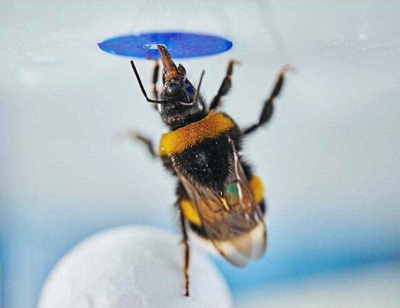
Problem-solving using novel solutions without explicit training is often considered a hallmark of cognitive flexibility. We investigated whether bumble bees (Bombus terrestris) could solve a novel object manipulation task spontaneously. Bees trained to associate a blue ring (“flower”) on the floor with a reward successfully moved a ball underneath a flower relocated to the ceiling to reach the flower. In control experiments in which the flower was out of sight when ball movement began and remained hidden during transport, bees still succeeded in the task. These results suggest that these were goal-directed actions rather than reinforcement-based associations driven by perceptual feedback. Our findings provide evidence that bumble bees can exhibit spontaneous problem-solving, challenging the notion that such advanced cognitive abilities are exclusive to large-brained vertebrates.