Study of more than 1,200 patients using donanemab finds cognitive benefits last at least three years
Animal models have long been central to neuroscience, providing direct experimental access to neural processes underlying perception, action, cognition, and disease. Over the past century, work in non-human primates (NHPs), rodents, and other species has established key principles of neural organization and behavior and has supported much of translational neuroscience. However, the institutional and material conditions that sustain animal-based research are now changing in fundamental ways. Ethical and regulatory requirements have intensified, costs and approval timelines have increased, and global supply chains, particularly for NHPs, have become fragile. In parallel, advances in human neuroscience, stem-cell-derived systems, and computational approaches have matured to the point that they challenge the historical reliance on animals for many classes of questions. These forces are not eliminating animal research, but they are reshaping the conditions under which it remains feasible, competitive, and scientifically justified. In this Perspective, we examine how these converging pressures are reconfiguring animal-based neuroscience. We review long-term trends in animal use and accessibility, highlighting species-specific constraints and emerging geopolitical asymmetries. We then analyze the growing role of alternative and complementary platforms, including human brain organoids, genetically engineered rodents, small primates, and ‘human-centric’ neurophysiological and imaging approaches, emphasizing both their strengths and limitations. Finally, we discuss the implications of this diversification for research planning, training, and scientific organization. We argue that the future of neuroscience will be defined not by the disappearance of animal models, but by their integration into hybrid experimental frameworks that preserve mechanistic rigor while adapting to evolving scientific and societal constraints.
Keywords: animal models; neuroscience methodology; alternative experimental platforms; translational validity; research ethics and regulation.
“IWMT and Human Consciousness Hypothesis”
A talk by Adam Safron, Victoria Klimaj and Zahra Sheikhbahaee recorded live at the AAAI 2026 conference.
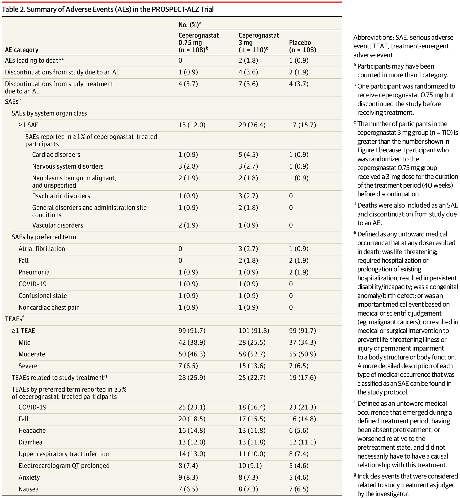
This multicenter, randomized, double-blind, placebo-controlled, phase 2 trial of ceperognastat in participants with early symptomatic AD was conducted at 72 centers in Australia, Canada, Japan, Poland, and the US from September 2021 to July 2025. The study adhered to the ethical principles outlined in the Declaration of Helsinki20 and other international guidelines. All participants provided written, informed consent before any study-related procedures. The study followed the Consolidated Standards of Reporting Trials (CONSORT) reporting guideline.21 The protocol was approved by an ethics committee at each participating center, and unblinded safety data were reviewed approximately every 3 months by an independent unblinded external data and safety monitoring committee. Participants were compensated for study participation. The study protocol and statistical analysis plan are provided in Supplements 1 and 2, respectively.
Eligible participants were aged 60 to 85 years and had a Mini-Mental State Examination (MMSE) score of 22 to 30, Clinical Dementia Rating Scale (CDR) Global Score of 0.5 or 1 with a memory box score greater than or equal to 0.5, elevated plasma level of tau phosphorylated at residue 217 (p-tau217), and evidence of elevated brain tau levels by flortaucipir F18 positron emission tomography (PET) scan at the time of screening. Demographic information, including race and ethnicity, was collected to allow for characterization of potential differences in treatment effects by demographic characteristic. Race and ethnicity were self-reported by participants based on fixed categories. All study participants and study staff were blinded to treatment assignment during the treatment phase.
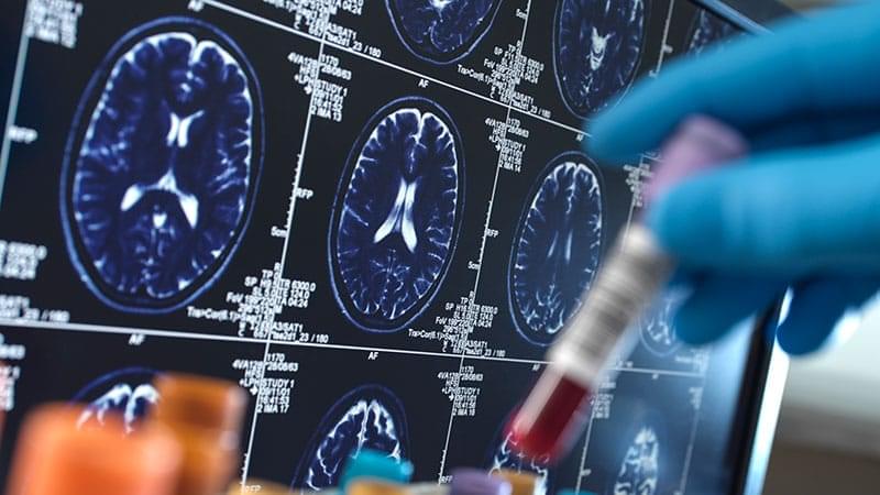
LONDON — Blood-based biomarker (BBM) testing may enable primary care physicians (PCPs) to diagnose Alzheimer’s disease (AD) as accurately as dementia specialists, potentially expanding access to accurate diagnosis beyond memory clinics, new research suggests.
In a prospective study of more than 1,300 patients, PCPs achieved 93% diagnostic accuracy after reviewing BBM results, which was comparable to the 94% accuracy of dementia specialists. The test also changed clinicians’ diagnoses and management plans in a substantial proportion of cases.
“By equipping primary care practitioners with blood test results, we see that they’re as accurate as dementia experts in definitely ruling out [AD],” study investigator Sebastian Palmqvist, MD, PhD, senior consultant neurologist and associate professor, Lund University, Lund, Sweden, told Medscape Medical News.
Cancers don’t come much worse than the brain cancer glioblastoma, and it is notoriously difficult to treat. Even with surgery, radiation, and chemotherapy, fewer than 30 percent of patients are alive two years after diagnosis.
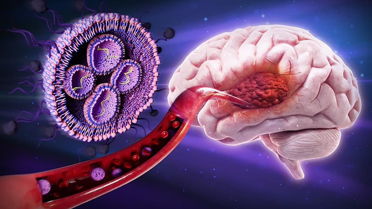
Scientists are busy hunting for treatment approaches that can improve those survival rates, and a team from Oregon State University has now found a potential new angle for attacking these tumors: sugar-coated nanoparticles.
As detailed in a mouse study published in the Journal of Controlled Release, the sugar ‘disguise’ used by the nanoparticles helps them cross the blood-brain barrier to the site of cancer, while also directly targeting glioblastoma and avoiding measurable toxicity in major organs.
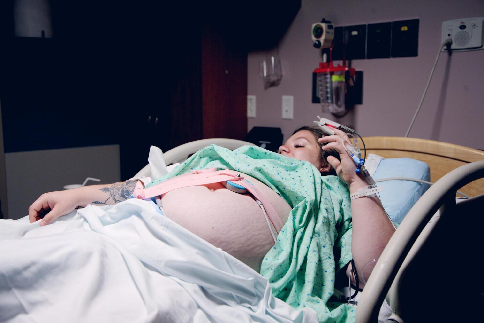
Having an epidural during labor is not associated with clinically significant increased risks of harm to newborn babies, including brain injury, severe breathing problems, sepsis and death, or cerebral palsy later in childhood, according to a study published in The BMJ.
The researchers say these findings “support widening availability and equitable access to epidural analgesia as a safe component of intrapartum care.”
Epidural analgesia in labor provides effective pain relief and may help reduce complications in mothers after giving birth, but evidence of its effect on newborn and child health is limited.
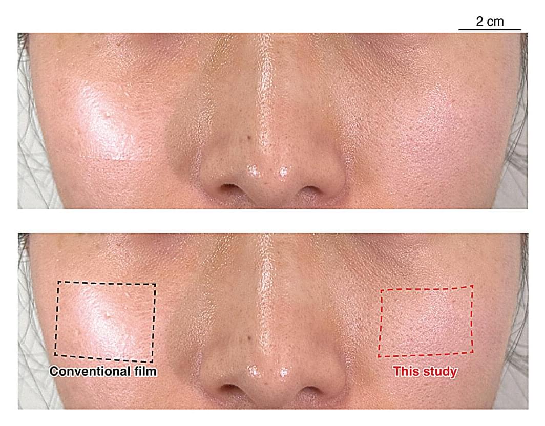
While wearable health sensors are becoming increasingly common, current iterations are awkward to wear. For example, devices attached to the face can draw unwanted attention, increase self-consciousness and influence the signals users are trying to measure. However, recent research may have found a solution by introducing ultrathin sensors that cannot be seen by observers or felt by the wearer.
In an article published in Science Advances, researchers from the Institute of Industrial Science, The University of Tokyo, and collaborating institutions reported developing thin, stretchable on-skin electrodes that are effectively invisible when worn on the face. The new technology can measure biological signals while remaining undetectable by eye and touch, allowing monitoring to take place under more natural conditions.
Biosignals such as eye movements, facial muscle activity and brain activity provide valuable information for health care monitoring and human-machine interaction. However, conventional facial electrodes can alter a person’s appearance and affect social interactions, creating what are called appearance artifacts—changes in behavior or psychological state caused simply by wearing a device that the individual and others can see.
