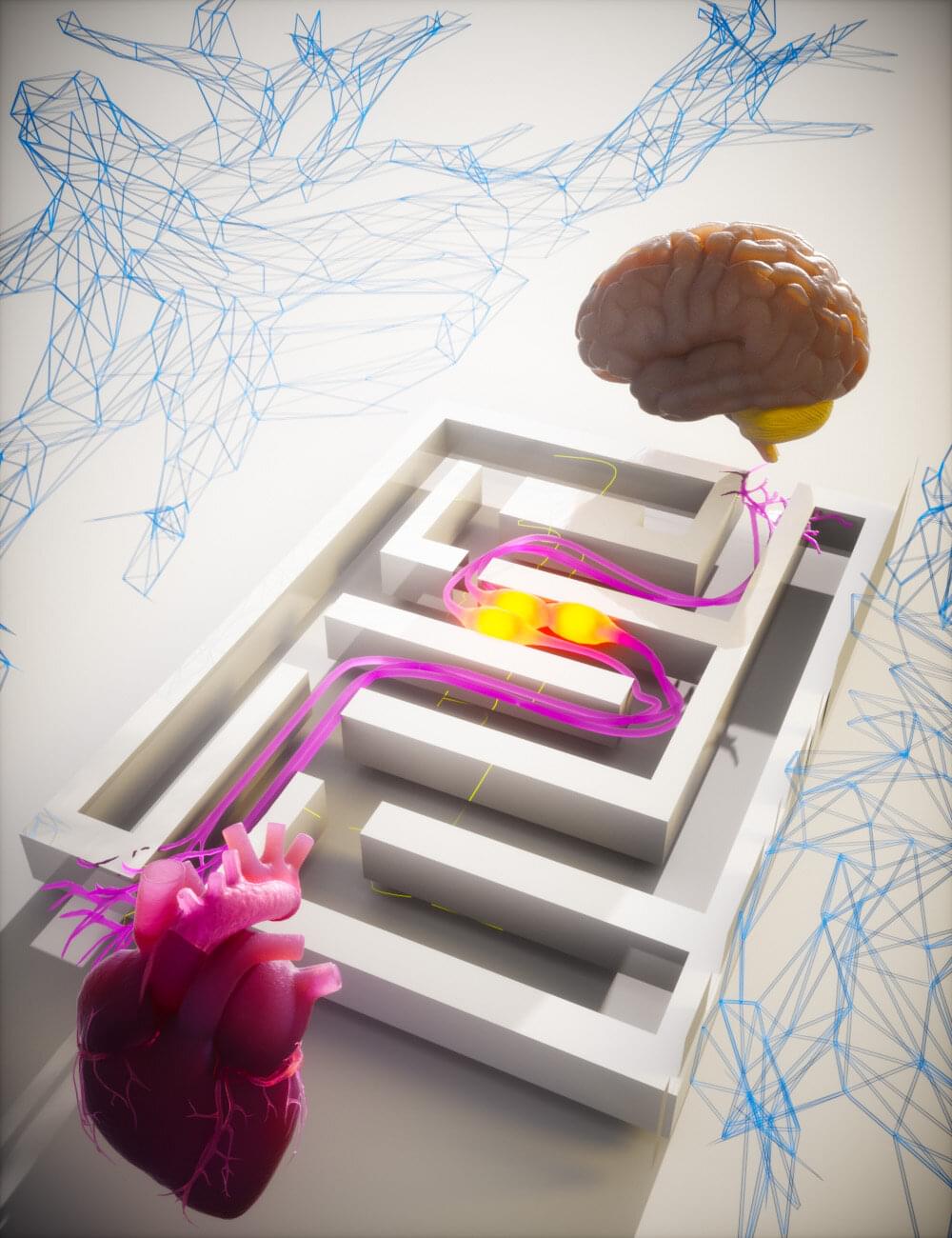
Using advanced microscopes that capture brain cell anatomy and activity, a portion of a mouse’s brain was mapped and rendered into a 3D atlas that creates new possibilities for neuroscience.
Category: neuroscience – Page 3
When it comes to early detection of cognitive impairment, a new study suggests that the nose knows. Researchers from Mass General Brigham have developed olfactory tests—in which participants sniff odor labels that have been placed on a card—to assess people’s ability to discriminate, identify and remember odors. They found that participants could successfully take the test at home and that older adults with cognitive impairment scored lower on the test than cognitively normal adults.
Results are published in Scientific Reports.
“Early detection of cognitive impairment could help us identify people who are at risk of Alzheimer’s disease and intervene years before memory symptoms begin,” said senior author Mark Albers, MD, Ph.D., of the Laboratory of Olfactory Neurotranslation, the McCance Center for Brain Health, and Department of Neurology at Massachusetts General Hospital.
Neurobehavioural disturbances such as loss of motivation have profound effects on the lives of many people living with Parkinson’s disease (PD), as well as other brain disorders. The field of decision-making neuroscience, underpinned by a plethora of work across species, provides an important framework within which to investigate apathy in clinical populations. Here we review how changes in a number of different processes underlying value-based decision making may lead to the common phenotype of apathy in PD. The application of computational models to probe both behaviour and neurophysiology show promise in elucidating these cognitive processes crucial for motivated behaviour. However, observations from the clinical management of PD demand an expanded view of this relationship, which we aim to delineate. Ultimately, effective treatment of apathy may depend on identifying the pattern in which decision making and related mechanisms have been disrupted in individuals living with PD.
For decades, scientists assumed that neural stem cells (NSCs) only occur in the brain and spinal cord. A new international study, led by Hans Schöler of the Max Planck Institute for Molecular Biomedicine in Münster, has now refuted this assumption and discovered a new type of neural stem cell outside the central nervous system (CNS) that opens up enormous possibilities for the development of therapies for neurological diseases. The study is published in the journal Nature Cell Biology.
In 2014, an article titled “Stimulus-triggered fate conversion of somatic cells into pluripotency” was published in Nature. This publication initially caused quite a stir because it opened up a simple way to obtain pluripotent stem cells. The induction of pluripotent stem cells without the need for viral vectors, as Shinya Yamanaka had done and for which he received the Nobel Prize, would have been too good to be true.
Although the laboratory of Schöler at the Max Planck Institute for Molecular Biomedicine, like many others, tried to repeat the experiment that described the “stimulus-triggered acquisition of pluripotency” (STAP) based on treating somatic cells with low pH. However, the generation of pluripotent cells failed regardless of the culture conditions and tissues used—and the corresponding paper was eventually retracted several months after publication.
When doing tasks that require little attention, people who let their minds wander show brain activity similar to sleep. This sleep-like activity has been linked to improved performance. When people let their minds wander during tasks that require focus and active thinking, their learning and perf
Trying to grasp the brain’s complexity is a little like trying to comprehend the vastness of space – it feels way beyond our scope of understanding.
By mapping a small part of a mouse brain down to an amazing level of detail, new research could help us grasp the magnitude of the neurological cosmos inside our heads.
Though the volume of brain matter analyzed was barely the size of a grain of sand, the researchers still had to describe the relationships between 84,000 neurons via half a billion synapse connections and 5.4 kilometers (3.4 miles) of neural wiring.
Previous studies have suggested that microRNAs are critical for brain development, but their specific role in differentiation—the process of stem cells maturing into specialized cells—remained unclear.
“When neurons develop, they need to at some point decide what subtype they will become, but we really didn’t know much about the blueprint that instructs this differentiation,” says the author. “There was a lot of evidence suggesting that microRNAs might have a very important role here, but because the tools were not good enough, we couldn’t really nail down that question until now.”
The team focused on Purkinje cells, which comprise less than 1% of cells in the cerebellum. Purkinje cells integrate information from different parts of the brain and body, enabling us to make smooth, controlled movements. They are some of the largest brain cells and have a tree-like appearance—a single axon “trunk” that supports a system of “branches” known as the dendritic arbor. Purkinje cells are also surrounded by structures called climbing fibers that wrap around the cells’ dendrites and deliver information from other parts of the brain.
To attain their large size and elaborate arbor, Purkinje cell development involves prolonged periods of growth and branching. In mice, the long process of Purkinje cell development is complete around four weeks after birth.
To investigate how microRNAs are involved in neuron differentiation, the team developed new tools that temporarily turn off microRNA function during specific developmental windows. They found that microRNAs are critical during two phases in Purkinje cell development: inhibiting microRNAs during the first week after birth resulted in Purkinje cells with less complex dendritic arbors and smaller cerebellums. In contrast, inhibiting microRNAs during the third week after birth prevented the Purkinje cells from forming synaptic connections with climbing fibers. These findings shed light on how microRNAs control the precise timing of different aspects of Purkinje cell development that were previously thought to happen concurrently.
The team also developed a mouse model to identify which genes the microRNA molecules were targeting. Using this system, they identified two microRNAs critical for Purkinje cell development (miR-206 and miR-133) and four gene targets (Shank3, Prag1, Vash1, and En2). When they compared the Purkinje cell microRNA-target map to a map for pyramidal neurons—a functionally different but similar-looking brain cell—they showed that the two cell types follow very different microRNA blueprints during development.
MicroRNAs (miRNAs) serve as key regulators of enteric nervous system development, orchestrating migration, proliferation, and differentiation of enteric nervous system progenitors.
Aberrant miRNA expression underpins the pathogenesis of several enteric neuropathies, including Hirschsprung’s disease.
A convergence of miRNA activity across distinct enteric neuropathies highlights shared molecular pathways, exemplified by the miR-200 family.
Modulating the expression of miRNAs to influence their associated gene expression networks has therapeutic potential for enteric neuropathies. https://sciencemission.com/MicroRNA-regulation-of-enteric-ne…nd-disease
The enteric nervous system (ENS), an elaborate network of neurons and glia woven through the gastrointestinal tract, is integral for digestive physiology and broader human health. Commensurate with its importance, ENS dysfunction is linked to a range of debilitating gastrointestinal disorders. MicroRNAs (miRNAs), with their pleiotropic roles in post-transcriptional gene regulation, serve as key developmental effectors within the ENS. Herein, we review the regulatory dynamics of miRNAs in ENS ontogeny, showcasing specific miRNAs implicated in both congenital and acquired enteric neuropathies, such as Hirschsprung’s disease (HSCR), achalasia, intestinal neuronal dysplasia (IND), chronic intestinal pseudo-obstruction (CIPO), and slow transit constipation (STC).
In what is considered the most complicated neuroscience experiment ever attempted, scientists from the Allen Institute and global collaborators have created the largest, most complex wiring diagram and functional map of the brain to date.