University of Virginia Comprehensive Cancer Center scientists have developed a promising new experimental approach to targeting glioblastoma, the most common and deadliest brain cancer. The approach could overcome many of the limitations of treatments using existing drugs.
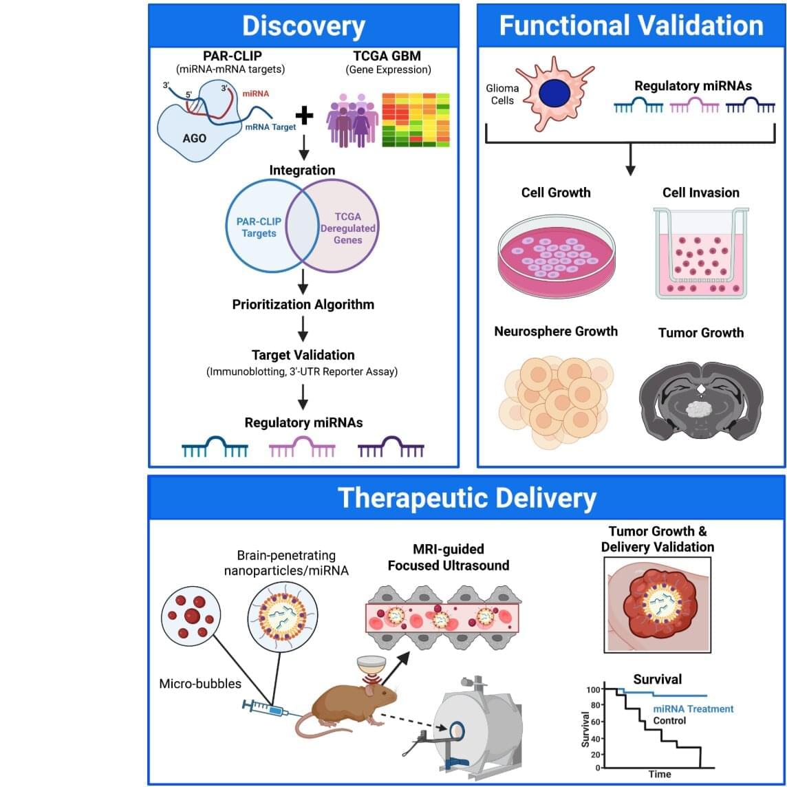
UVA’s Roger Abounader, MD, Ph.D., and colleagues have identified “microRNAs” that can simultaneously suppress multiple malfunctioning genes responsible for glioblastoma’s formation and growth. The scientists use a combination of brain-penetrating nanoparticles, focused ultrasound waves and microbubbles to deliver the miRNAs through the brain’s natural protective barrier—a barrier that typically blocks treatments for tumors and neurodegenerative diseases. The study is published in the Journal of Clinical Investigation.
“This new approach could help target numerous molecules that promote cancer growth, including those for which no drugs exist, at the same time to achieve better therapies,” said Abounader, a professor at UVA’s School of Medicine, Department of Microbiology, Immunology and Cancer Biology, Comprehensive Cancer Center and Center for RNA Science and Medicine. “We are hoping to translate our findings into future clinical trials for patients with glioblastoma and other brain tumors.”