Most everyday moments are quickly forgotten, while others stay with us for a lifetime. How the brain determines which memories are retained and which are lost remains an open question.
A neuroscience team at the University of Tübingen, led by Professor Andrea Burgalossi of the Institute of Neurobiology and the Werner Reichardt Center for Integrative Neuroscience (CIN), has found that spontaneous fluctuations in the brain’s internal state influence how ready the memory system is to store new information. The new study has been published in Nature Communications.
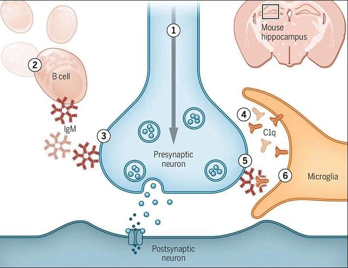
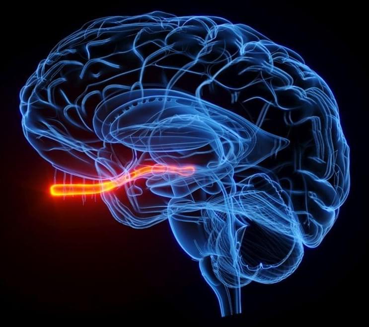
The researchers used mice to investigate the neural mechanisms of memory formation, focusing on the hippocampus, a region of the brain known to be essential to the formation of episodic memories. The hippocampus of mammals, including mice and humans, contains neurons called place cells that represent experiences in the brain.