Category: neuroscience – Page 2

Research progress on 40 Hz sensory stimulation for the treatment of Alzheimer’s disease
Abstract:
Alzheimer’s disease (AD) is a prevalent neurodegenerative disorder characterized by β-amyloid (Aβ) deposition, tau protein hyperphosphorylation, and synaptic dysfunction. In recent years, 40 Hz sensory stimulation—including visual, auditory, and multimodal modalities—has emerged as a novel, non-invasive intervention demonstrating potential efficacy in both animal models and preliminary clinical studies. Preclinical evidence indicates that such stimulation can markedly reduce cerebral Aβ burden (by approximately 37%–53%), inhibit tau protein phosphorylation, enhance neuronal network synchrony and synaptic plasticity, and improve learning and memory performance. Limited human trials suggest that 40 Hz sensory stimulation is safe and well tolerated in patients with mild cognitive impairment (MCI) and early-stage AD, with a slowing trend in cognitive scale score decline following intervention. This review summarizes the mechanisms of action, experimental evidence from animal models, and advances in clinical application of 40 Hz sensory stimulation in AD prevention and treatment. It further explores the potential for multimodal combination therapies integrating sensory stimulation with cognitive training, pharmacological interventions, and lifestyle modifications, and addresses challenges such as optimal timing of intervention and the influence of ambient electromagnetic fields in real-world settings. Current evidence supports 40 Hz sensory stimulation as a feasible, multi-target, and safe adjunctive intervention; however, its efficacy and applicability must be verified through multicenter, randomized controlled trials with long-term follow-up.

Multifunctional Organic Materials, Devices, and Mechanisms for Neuroscience, Neuromorphic Computing, and Bioelectronics
Neuromorphic computing has the potential to overcome limitations of traditional silicon technology in machine learning tasks. Recent advancements in large crossbar arrays and silicon-based asynchronous spiking neural networks have led to promising neuromorphic systems. However, developing compact parallel computing technology for integrating artificial neural networks into traditional hardware remains a challenge. Organic computational materials offer affordable, biocompatible neuromorphic devices with exceptional adjustability and energy-efficient switching. Here, the review investigates the advancements made in the development of organic neuromorphic devices. This review explores resistive switching mechanisms such as interface-regulated filament growth, molecular-electronic dynamics, nanowire-confined filament growth, and vacancy-assisted ion migration, while proposing methodologies to enhance state retention and conductance adjustment. The survey examines the challenges faced in implementing low-power neuromorphic computing, e.g., reducing device size and improving switching time. The review analyses the potential of these materials in adjustable, flexible, and low-power consumption applications, viz. biohybrid spiking circuits interacting with biological systems, systems that respond to specific events, robotics, intelligent agents, neuromorphic computing, neuromorphic bioelectronics, neuroscience, and other applications, and prospects of this technology.
Keywords: Brain-inspired neuromorphic computing; Neuromorphic bioelectronics; Neuroscience; Organic materials; Resistive switching mechanisms.
© 2025. The Author(s).

Neuromorphic Sentiment Analysis Using Spiking Neural Networks
Over the past decade, the artificial neural networks domain has seen a considerable embracement of deep neural networks among many applications. However, deep neural networks are typically computationally complex and consume high power, hindering their applicability for resource-constrained applications, such as self-driving vehicles, drones, and robotics. Spiking neural networks, often employed to bridge the gap between machine learning and neuroscience fields, are considered a promising solution for resource-constrained applications. Since deploying spiking neural networks on traditional von-Newman architectures requires significant processing time and high power, typically, neuromorphic hardware is created to execute spiking neural networks. The objective of neuromorphic devices is to mimic the distinctive functionalities of the human brain in terms of energy efficiency, computational power, and robust learning. Furthermore, natural language processing, a machine learning technique, has been widely utilized to aid machines in comprehending human language. However, natural language processing techniques cannot also be deployed efficiently on traditional computing platforms. In this research work, we strive to enhance the natural language processing traits/abilities by harnessing and integrating the SNNs traits, as well as deploying the integrated solution on neuromorphic hardware, efficiently and effectively. To facilitate this endeavor, we propose a novel, unique, and efficient sentiment analysis model created using a large-scale SNN model on SpiNNaker neuromorphic hardware that responds to user inputs. SpiNNaker neuromorphic hardware typically can simulate large spiking neural networks in real time and consumes low power. We initially create an artificial neural networks model, and then train the model using an Internet Movie Database (IMDB) dataset. Next, the pre-trained artificial neural networks model is converted into our proposed spiking neural networks model, called a spiking sentiment analysis (SSA) model. Our SSA model using SpiNNaker, called SSA-SpiNNaker, is created in such a way to respond to user inputs with a positive or negative response. Our proposed SSA-SpiNNaker model achieves 100% accuracy and only consumes 3,970 Joules of energy, while processing around 10,000 words and predicting a positive/negative review. Our experimental results and analysis demonstrate that by leveraging the parallel and distributed capabilities of SpiNNaker, our proposed SSA-SpiNNaker model achieves better performance compared to artificial neural networks models. Our investigation into existing works revealed that no similar models exist in the published literature, demonstrating the uniqueness of our proposed model. Our proposed work would offer a synergy between SNNs and NLP within the neuromorphic computing domain, in order to address many challenges in this domain, including computational complexity and power consumption. Our proposed model would not only enhance the capabilities of sentiment analysis but also contribute to the advancement of brain-inspired computing. Our proposed model could be utilized in other resource-constrained and low-power applications, such as robotics, autonomous, and smart systems.
Keywords: SpiNNaker; artificial neural network; natural language processing; neuromorphic computing; sentiment analysis; spiking neural networks.
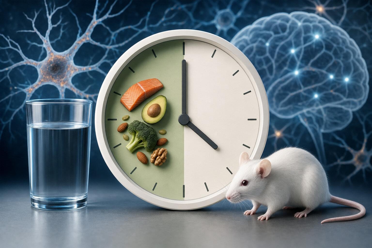
How intermittent fasting may shield the brain from chronic stress
Chronic stress, the prolonged exposure to psychological and/or physical strain, is known to be a risk factor for depression, anxiety and some other psychiatric disorders. Past studies suggest that chronic stress disrupts the integrity of myelin, a fatty insulating layer that surrounds nerve fibers and helps electrical signals travel efficiently between brain cells.
Identifying lifestyle changes that can reverse or diminish the adverse effects of chronic stress on the brain could be advantageous, as they could potentially help prevent or delay the onset of various psychiatric conditions. Recently, some researchers have been exploring the potential brain benefits of intermittent fasting (IF), a dietary pattern that entails alternating between set periods of eating and fasting.
Past findings suggest that IF can improve people’s metabolism and help reduce inflammation, the body’s natural response to disease or injury. Yet its effects on people’s mental health and well-being have not yet been clearly determined.
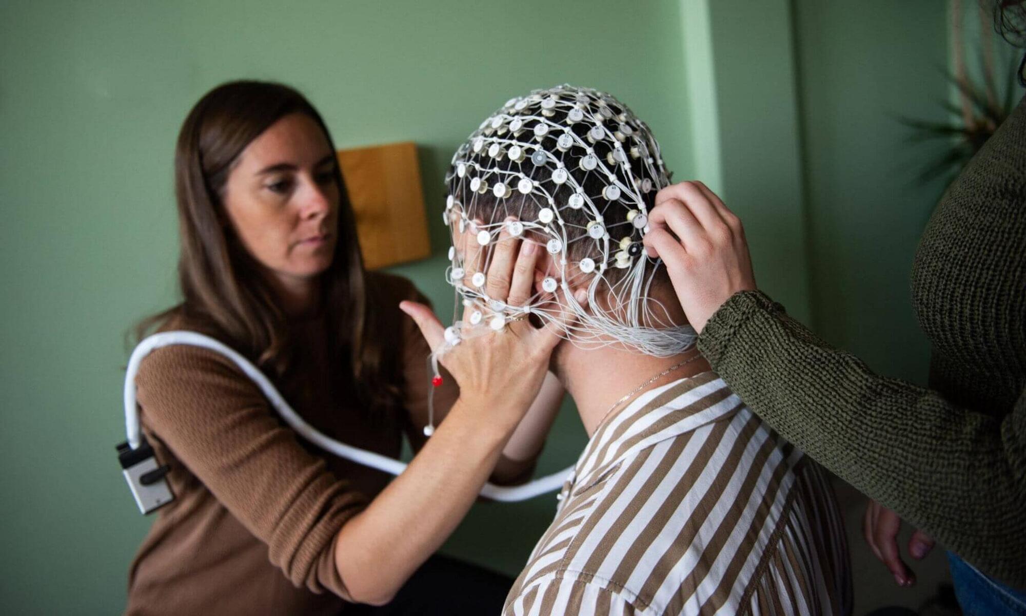
‘Pink noise’ can help make anesthesia work better during surgery
In the brain, specific electrical waves are associated with different states of consciousness. For instance, delta waves—also known as slow waves—are especially prevalent during deep sleep, as well as during states of unconsciousness induced by coma and general anesthesia. They are considered a “signature” of these altered states of consciousness.
Over a decade ago, research showed that it is possible to amplify these delta waves through highly precise auditory stimulation, a technique initially studied in the context of sleep.
Now researchers at Université de Montréal are bringing this technique into the operating room to help optimize general anesthesia, which also induces a state characterized by abundant delta waves.
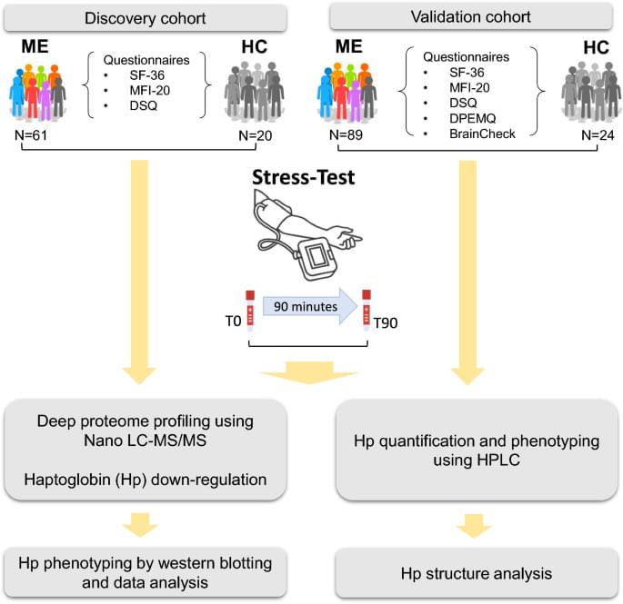
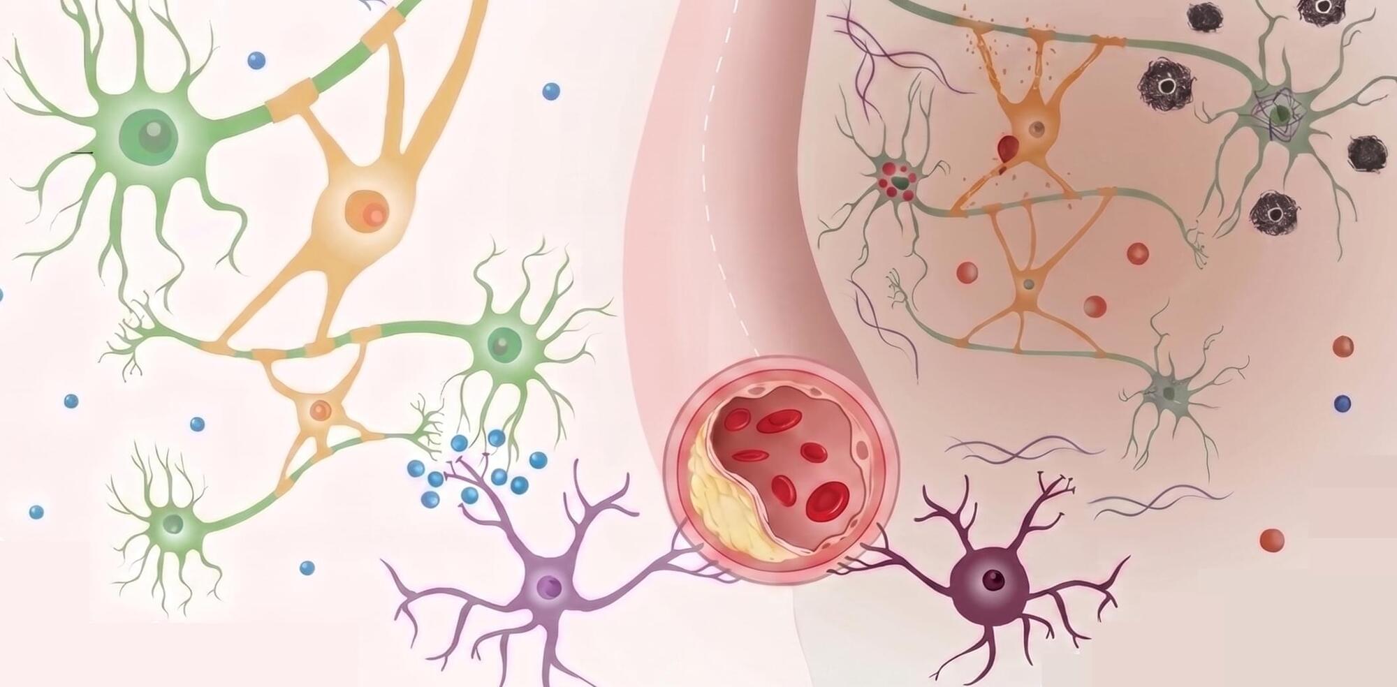
Haptoglobin phenotypes and structural variants associate with post-exertional malaise and cognitive dysfunction in myalgic encephalomyelitis
Myalgic encephalomyelitis (ME) is a chronic, multisystem illness characterized by post-exertional malaise (PEM) and cognitive dysfunction, yet the molecular mechanisms driving these hallmark symptoms remain unclear. This study investigated haptoglobin (Hp) as a potential biomarker of PEM severity and cognitive impairment in ME, with a focus on Hp phenotypes and structural proteoforms.
A longitudinal case–control study was conducted in 140 ME patients and 44 matched sedentary healthy controls. In the discovery phase, global plasma proteomic profiling was performed in 61 ME patients and 20 controls before and after a standardized, non-invasive stress protocol in order to induce PEM. Associations between Hp levels, phenotype, and cognitive performance were assessed. In the validation phase, plasma Hp concentrations and proteoform composition were analyzed in an independent cohort of 89 ME patients and 24 controls using high-performance liquid chromatography (HPLC).
ME patients demonstrated a significant reduction in Hp levels following post-exertional stress. Lower baseline Hp concentrations were associated with impaired cognitive performance. Hp phenotypes were differentially associated with symptom burden, with the Hp2-1 phenotype enriched in ME and linked to greater PEM severity and cognitive deficits compared to Hp1-1 and Hp2-2. HPLC analysis revealed altered Hp proteoform profiles in the Hp2-1 subgroup, including increased high-mass tetrameric and pentameric forms and shorter retention times indicative of structural changes. In contrast, the Hp1-1 phenotype was associated with milder symptoms and greater cognitive resilience.
Brain aneurysm map reveals cell types tied to rupture risk
A new study from UC San Francisco shows how certain cells in the brain may cause aneurysms to weaken and rupture. It helps explain why some aneurysms burst while others do not and could lead to new ways of predicting and possibly preventing strokes.
Brain aneurysms are bulges in blood vessels that can go unnoticed for years. If they rupture, they can cause a severe and often deadly type of stroke. About one in 50 Americans has a brain aneurysm, but doctors still struggle to predict which ones are most dangerous.
The new study helps to unpack the biology behind these events by mapping the cells in artery walls and the interactions that weaken them.