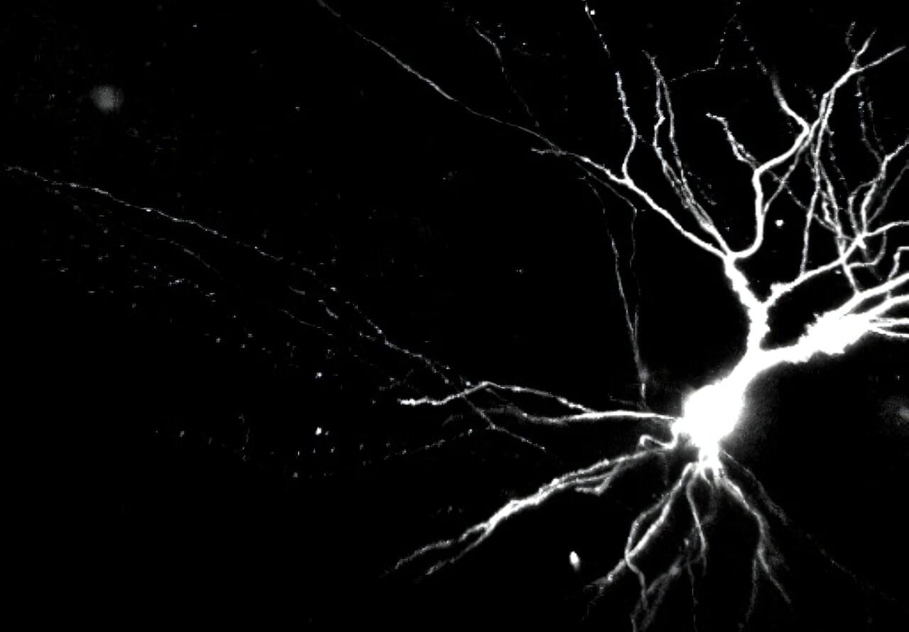
Toggle light / dark theme

A new study from Columbia University Mailman School of Public Health and the Columbia Butler Aging Center suggests that risk factors and biomarkers related to Alzheimer’s disease are associated with cognition much earlier in life than previously recognized. The study highlights significant associations between cognition and Alzheimer’s disease risk factors as young as ages 24 to 44 and underscores the importance of early prevention.

This is the first study to systematically examine Alzheimer’s disease risk factors, including biomarkers related to in a large group of generally healthy middle-aged individuals in the U.S. The findings are published in The Lancet-Regional Health Americas.

“Previously, research on Alzheimer’s disease risk factors has focused on individuals aged 50 and older,” said Allison Aiello, Ph.D., James S. Jackson Healthy Longevity Professor of Epidemiology in the Butler Aging Center and Columbia Mailman School. “The potential impact of our findings is substantial, offering clinicians and health researchers a clearer understanding of the early emergence of Alzheimer’s disease risk factors and their association with cognition before middle age.

There are several physiological reasons why biological organisms sleep. One key one concerns brain metabolism. In our article we discuss the role of metabolism in myelin, based on the recent discovery that myelin contains mitochondrial components that enable the production of adenosine triphosphate (ATP) via oxidative phosphorylation (OXPHOS). These mitochondrial components in myelin probably originate from vesiculation of the mitochondrial membranes in form from mitochondrial derived vesicles (MDVs). We hypothesize that myelin acts as a proton capacitor, accumulating energy in the form of protons during sleep and converting it to ATP via OXPHOS during wakefulness. Empirical evidence supporting our hypothesis is discussed, including data on myelin metabolic activity, MDVs, and allometric scaling between white matter volume and sleep duration in mammals.

A new way to deliver disease-fighting proteins throughout the brain may improve the treatment of Alzheimer’s disease and other neurological disorders, according to University of California, Irvine scientists. By engineering human immune cells called microglia, the researchers have created living cellular “couriers” capable of responding to brain pathology and releasing therapeutic agents exactly where needed.

The study, published in Cell Stem Cell, demonstrates for the first time that derived from induced pluripotent stem cells can be genetically programmed to detect disease-specific brain changes—like in Alzheimer’s disease—and then release enzymes that help break down those toxic proteins. As a result, the cells were able to reduce inflammation, preserve neurons and synaptic connections, and reverse multiple other hallmarks of neurodegeneration in mice.

For patients and families grappling with Alzheimer’s and related diseases, the findings offer a hopeful glimpse at a future in which microglial-based cell therapies could precisely and safely counteract the ravages of neurodegeneration.

Results of a randomized, controlled clinical trial in Japan among more than 170 children aged 1 to 6 who underwent surgery show that by using EEG readings of brain waves to monitor unconsciousness, an anesthesiologist can significantly reduce the amount of the anesthesia administered to safely induce and sustain each patient’s anesthetized state.

On average, the patients experienced significant improvements in several post-operative outcomes, including quicker recovery and reduced incidence of delirium.

“I think the main takeaway is that in kids, using the EEG, we can reduce the amount of anesthesia we give them and maintain the same level of unconsciousness,” said study co-author Emery N. Brown, Edward Hood Taplin Professor of Medical Engineering and Computational Neuroscience at MIT and an anesthesiologist at Massachusetts General Hospital. The study appears in JAMA Pediatrics.

Miniature zombies are all around us, scuttling through the underbrush or flying through the air in nearly every continent on Earth. In Brazil, a fungus takes over ant brains, altering their circadian rhythms and social behaviors. In England, a virus forces caterpillars to climb high into the canopy, then slowly liquefies their bodies, which drip onto the leaves below. In Indonesia, a parasitoid wasp uses specialized venom to alter a cockroach’s brain chemistry, turning it into the perfect host for her young.

In her new book, Rise of the Zombie Bugs, self-described professional science nerd Mindy Weisberger introduces readers to a menagerie of mind-controlling parasites, and the scientists who have devoted their lives to the study of these peculiar organisms. Through these vivid tales of creatures bizarre enough to rival any fictional beast, Weisberger offers readers a peek into the fields of evolution, ecology, neuroscience, and molecular biology. She shows that these topics exist beyond dim lecture halls and dry textbooks: “Science is everything and everywhere,” she said.

Vision is one of the most crucial human senses, yet more than 300 million people worldwide are at risk of vision loss due to various retinal diseases. While recent advancements in retinal disease treatments have successfully slowed disease progression, no effective therapy has been developed to restore already lost vision—until now.

KAIST researchers led by Professor Jinwoo Kim from the Department of Biological Sciences have successfully developed a novel drug to restore vision through retinal nerve regeneration. The research is published in the journal Nature Communications. The study was co-authored by Dr. Eun Jung Lee of Celliaz Inc. and Museong Kim, a Ph.D. candidate at KAIST, as joint first authors.

The research team successfully induced neural regeneration and vision recovery in a disease-model mouse by administering a compound that blocks the PROX1 (Prospero Homeobox 1) protein, which suppresses retinal regeneration. The effect lasted for more than six months.

Statistics suggest that the size of families in many countries is shrinking and a growing number of parents worldwide either willingly or unwillingly end up only having one child. While many psychology studies have explored the differences between individuals who have siblings and those who don’t, the effects of not having any brothers or sisters on people’s brains and behavior are not yet fully understood.

Past research has yielded varying and sometimes contradictory results, which sometimes hinted at negative effects of being an only child and other times highlighted its positive implications. In addition, these negative and positive effects were found to be inconsistent across studies, with some studies suggesting that only children tend to do better at school, are more pro-social and less problematic, while others showed the opposite.

Researchers at Tianjin Medical University General Hospital and other institutes in China recently carried out a study aimed at better understanding how being an only child affects people’s brain and behavior during adulthood. Their findings, published in Nature Human Behaviour, highlight specific patterns in the brain’s development and activity, as well as behavioral tendencies, that are commonly observed in adults who grew up without siblings.