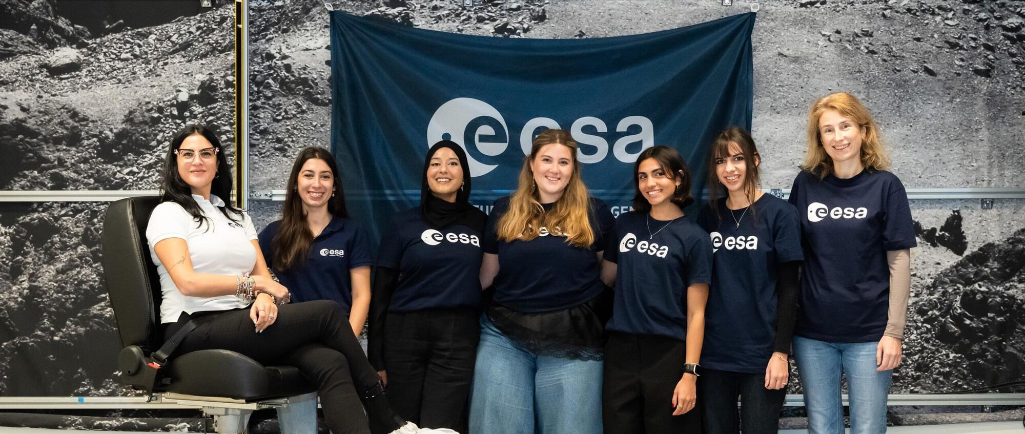
Toggle light / dark theme

Hormone supplementation in rhesus monkeys points to potential autism treatment

For years, Florida Tech’s Catherine Talbot, an assistant professor of psychology, has worked to understand the sociality of male rhesus monkeys and how low-social monkeys can serve as a model for humans with autism. Her most recent findings show that replenishing a deficient hormone, vasopressin, helped the monkeys become more social without increasing their aggression—a discovery that could change autism treatment.

Currently, the Centers for Disease Control and Prevention reports that one in 36 children in the United States is affected by autism spectrum disorder (ASD). That’s an increase from one in 44 children reported in 2018. Two FDA-approved treatments currently exist, Talbot said, but they only address associated symptoms, not the root of ASD. The boost in both prevalence and awareness of the disorder prompts the following question: What is the cause?

Some are naturally low-social, meaning they demonstrate poor social cognitive skills, while others are highly social. Their individual variation in sociality is comparable to how human sociality varies, ranging from people we consider social butterflies to those who are not interested in social interactions, similar to some people diagnosed with ASD, Talbot said. Her goal has been to understand how variations in biology and behavior influence social cognition.

Brain’s ‘blue spot’ key to healthy aging, early Alzheimer’s detection

In a demographically diverse sample of healthy people, Cornell researchers found dramatic changes over the human lifespan in the brain’s “blue spot”—a tiny region involved in cognition and believed to be the first affected by neurodegenerative conditions including Alzheimer’s disease.

Using specialized MRI scans to measure the intensity of neuromelanin, a pigment that gives the locus coeruleus (LC) its , the research team observed an inverted U-shaped curve that peaked in later middle age before dropping off sharply, a finding that helps characterize healthy aging patterns.

Maintaining a stronger blue signal after age 60 was associated with better cognitive performance, according to the study involving 134 participants aged 19 to 86. Because of the participants’ diversity, including about 40% who were non-white, the researchers also discovered higher peaks among Black participants and women, groups known to be more susceptible to Alzheimer’s.

Nasal microbiome may help explain link between olfactory dysfunction and cognitive decline

As humans age, particularly after middle age, their brain functions, cognitive abilities and memory can deteriorate to varying degrees. Aging-related disorders marked by cognitive decline, particularly dementia, have become increasingly widespread over the past decades.

Estimates suggest that the number of individuals diagnosed with dementia could increase from 55 million in 2019 to around 139 million by 2050. Understanding the factors contributing to and devising methods to detect the first signs of dementia is thus of the utmost importance, as it could help to reliably pick up its emergence and plan therapeutic interventions accordingly.

In recent years, some studies have found a link between people’s ability to perceive and identify odors (i.e., olfactory function) and their cognitive abilities as . While the relationship between and cognitive decline is now well-documented, whether one causes the other or they are the result of similar aging-related or neurodegenerative mechanisms remains unclear.

Limitations of the use of the MP-RAGE to identify neural changes in the brain: recent cigarette smoking alters gray matter indices in the striatum

The magnetization-prepared rapid gradient-echo (MP-RAGE) T1-weighted high resolution structural MRI is a mainstay tool used to identify morphometric biomarkers of disease conditions, progression and treatment effects despite a critical limitation: the relaxation signal on which inferences are based is nearly indistinguishable for gray matter vs. blood flow (Lu et al., 2004; Wright et al., 2008). Thus, apparent reported morphometric findings might be at least partially related to transient changes in blood flow or other physiological signals.

Consistent with this technical limitation, using a standard analysis technique, voxel based morphometry (VBM), we recently reported that a single dose of a medication had “apparent” effects on T1-weighted MRIs (Franklin et al., 2013). Specifically, we observed medication-induced decreases in gray matter volume in the anterior cingulate and other regions that overlapped with changes in brain blood flow (perfusion). Similarly, others have shown effects of medication on T1-weighted scans that are likely transient. For example, acute levodopa administration altered gray matter indices on T1-weighted images in the midbrain (Salgado-Pineda et al., 2006). Further, in a well-controlled longitudinal VBM study of patients with attention deficit hyperactivity disorder (ADHD), Hoekzema et al.

/* */