Toggle light / dark theme

Astrocytes are critical for fear memory

The team used a mouse model to understand how fear learning as a mechanism takes place in the brain, how fear-related memories can be retrieved, and the contribution of neurons versus astrocytes to fear learning.

Using fluorescent activity sensors, the team watched astrocytes respond in real time as fear memories were formed and later retrieved. As those memories were extinguished, astrocyte activity diminished. When the researchers then selectively increased or suppressed the signals astrocytes send to neighboring neurons, the strength of fear memories shifted in parallel, demonstrating that astrocytes are not just passive bystanders, but active participants in shaping fear.

Change in astrocyte activity also influenced neural circuits. When the astrocyte activity was disrupted, neurons could no longer form normal fear-related activity patterns and effectively transmit information about appropriate defensive reactions to brain regions that help control defensive behavior. These findings challenge neuron-centric models of fear by showing that fear memories aren’t produced by neurons alone.

The impact of disrupting astrocytes rippled beyond the amygdala. The manipulations also influenced how fear signals were relayed to the prefrontal cortex, a brain region that is key for decision-making. This suggests that astrocytes not only influence encoding of fear memories by the amygdala, but also how the brain uses those memories to determine appropriate responses to fearful situations.

Knowing that astrocytes play a key role in the retrieval of fear memories will reshape therapeutic interventions for disorders driven by persistent fearful memories such as post-traumatic stress disorder, anxiety disorders and phobias, the author said. If astrocytes help determine whether fear memories are expressed or successfully extinguished, then targeting astrocyte-related pathways, rather than neural pathways, could eventually complement neuron-focused therapies.


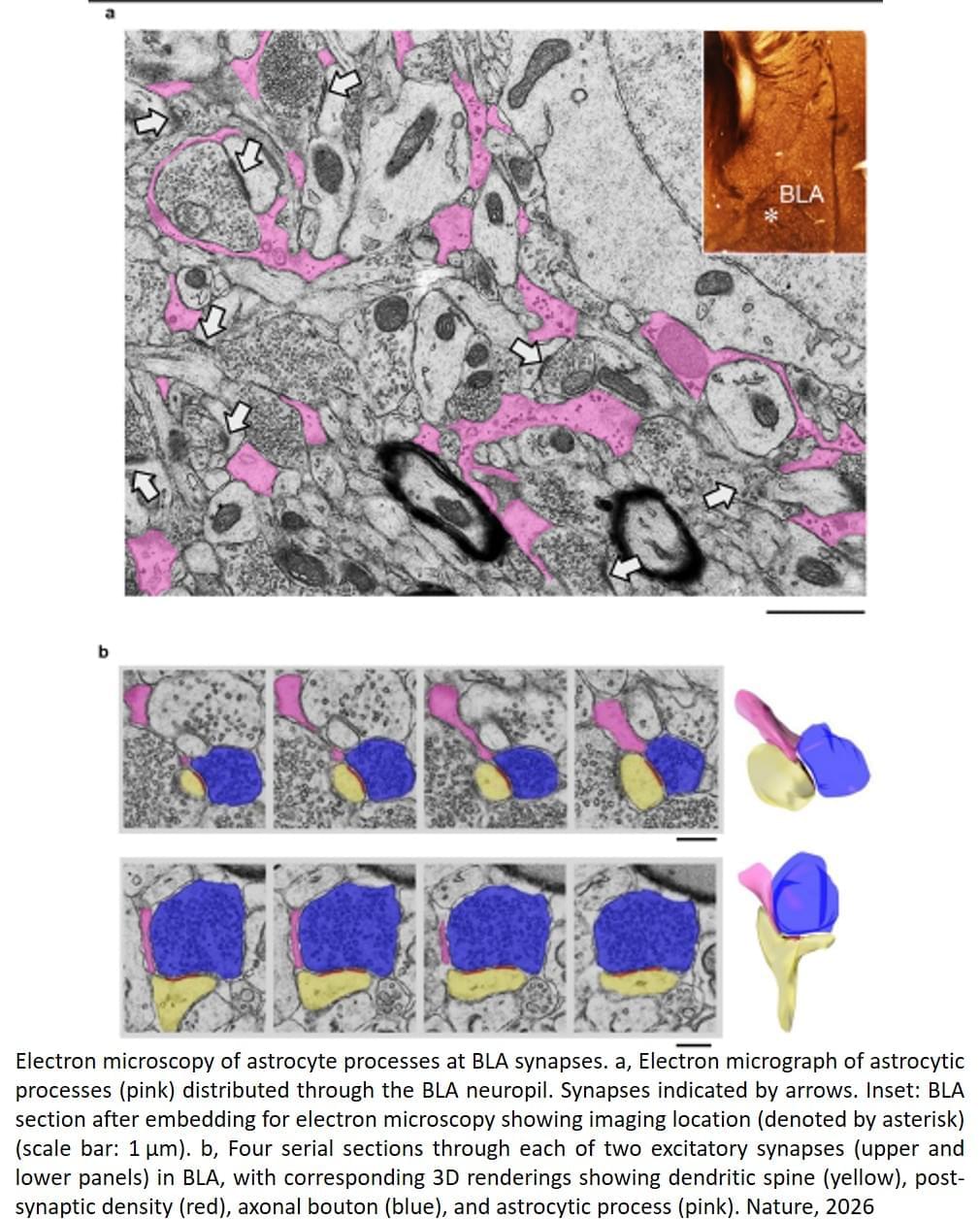
Picture a star-shaped cell in the brain, stretching its spindly arms out to cradle the neurons around it. That’s an astrocyte, and for a long time, scientists thought its job was caretaking the brain, gluing together neurons, and maintaining neural circuits.

Liquid Crystal Monomers Released from LCD Displays Accumulate in Endangered Marine Cetaceans Triggering Health Concerns

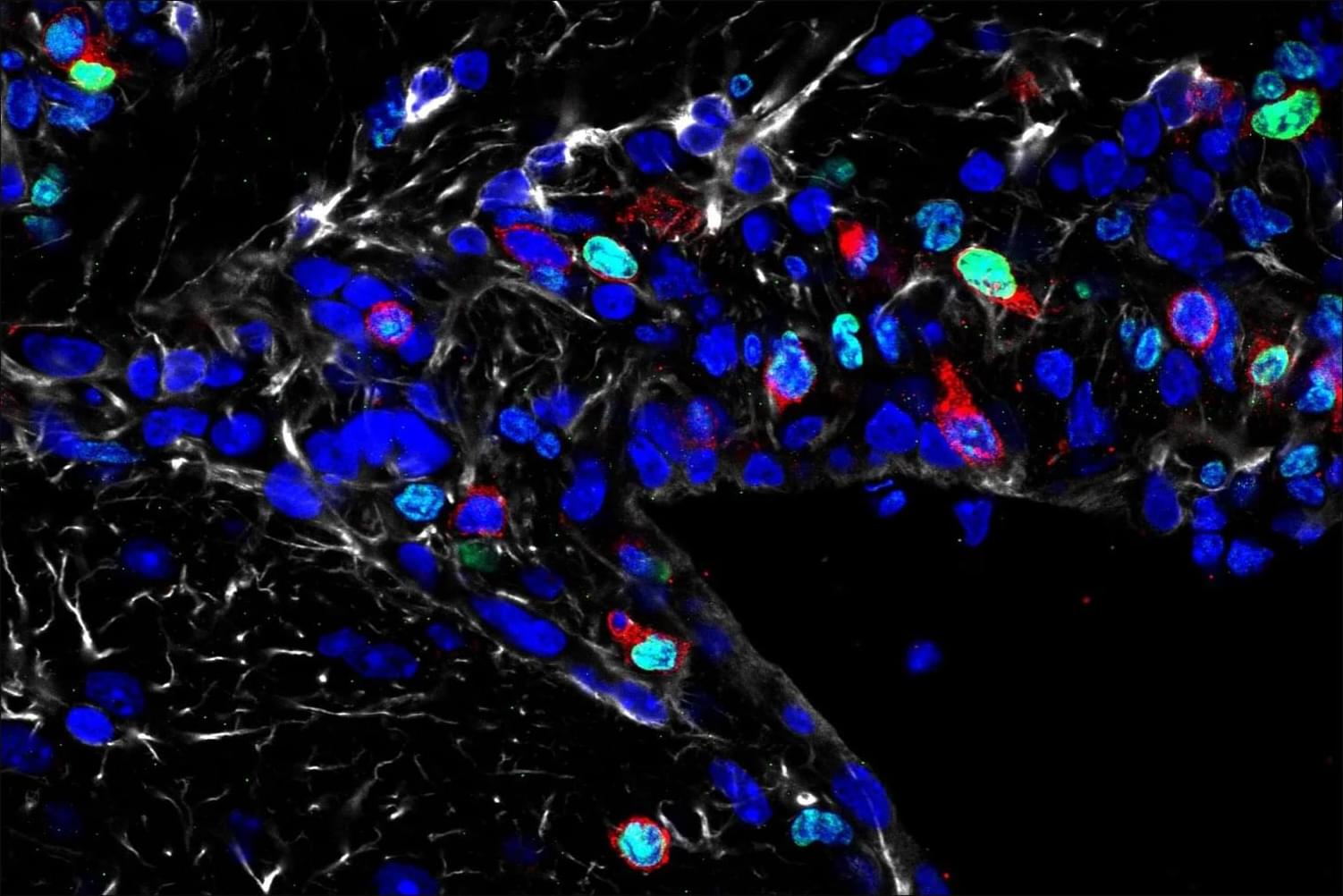
Liquid crystal monomers (LCMs), critical substances of liquid crystal displays in consumer electronics, are persistent pollutants, posing potential threats to marine ecosystems. Despite their bioaccumulative potential, their occurrence and possible biological impacts on marine megafauna remain understudied. We investigated LCM occurrence in Indo-Pacific humpback dolphins (Sousa chinensis) and finless porpoises (Neophocaena phocaenoides) collected from the South China Sea (2007–2021) and assessed their toxicity through in vitro assays using established dolphin cell lines. By employing robust source-tracing methodologies, we provide the first evidence that LCMs from household electronics and coastal e-waste accumulate in cetacean tissues, including blubber, muscle, and, critically, brain tissues, demonstrating blood–brain barrier penetration, a previously undocumented phenomenon of LCMs in mammalian wildlife. The temporal trend of LCM burden in porpoise blubber is correlated with shifts in global liquid crystal display production. Transcriptomic profiling revealed LCM-induced DNA damage, cell cycle arrest, and impaired cell division in cetacean cells. These findings suggest that LCMs may pose potential risks to the nervous system and other organs of marine mammals, warranting further investigation into their toxicological effects and possible implications for human health. By bridging critical gaps among everyday electronics, LCM contamination, and marine conservation, this study highlights the need for urgent regulatory actions and improved e-waste governance to mitigate ecological and public health risks.

Engineered immune therapy could help fight brain aging

Researchers at Stanford University engineered a modified version of the immune protein interleukin-10 (IL-10) that retains only its anti-inflammatory properties while eliminating its pro-inflammatory ones. When injected into aged mice, this modified protein stimulated the growth of new neurons and improved performance on memory and learning tasks, such as maze navigation and object recognition. The study, published in Immunity, suggests that age-related cognitive decline is linked to the accumulation of exhausted T-lymphocytes in the brain, chronic inflammation, and impaired microglial function — all of which reduce neurogenesis. The findings indicate that selectively modulating immune signaling could open new avenues for treating neurodegenerative diseases. The team plans to further investigate the protein’s mechanisms and explore ways to target specific cell types more precisely to minimize potential side effects.


A modified immune protein developed by Stanford researchers points to a novel strategy for combating age-related cognitive decline.

The Nervous System and Behavior

Many central issues with which neurosciences is concerned, such as how we perceive the world around us, how we learn from experience, how we remember, how we direct our movements, and how we communicate with each other, have commanded the attention of thoughtful men and women for centuries. But it was not until after World War II that neuroscience began to emerge as a separate and increasingly vigorous scientific discipline that has as its ultimate objective providing a satisfactory account of animal (including human) behavior in biological terms. This ambitious goal has as its basis the central realization that all behavior is, in the last analysis, a reflection of the function of the nervous system. It is the organized and coordinated activity of the nervous system that ultimately manifests itself in the behavior of the organism. The challenge to neuroscience then, is to explain, in physical and chemical terms, how the nervous system marshalls its signaling units to direct behavior.

The real magnitude of this challenge can perhaps be best judged by considering the structural and functional complexity of the human brain and the bewildering complexity of human behavior. The human brain is thought to be composed of about a hundred billion (1011) nerve cells and about 10 to 50 times that number of supporting elements or glial cells. Some nerve cells have relatively few connections with other neurons or with such effector organs as muscles or glands, but the great majority receive connections from thousands of other cells and may themselves connect with several hundred other neurons. This means that at a fairly conservative estimate the total number of functional connections (known as synapses) within the human brain is on the order of a hundred trillion (1014). But what is most important is that these connections are not random or indiscriminate:

They constitute the essential “wiring” of the nervous system on which the extraordinarily precise functioning of the brain depends. We owe to the great neuroanatomists of the last century, and especially to Ramón y Cajal, the brilliant insight that cells with basically similar properties are able to produce very different actions because they are connected to each other and to the sensory receptors and effector organs of the body in different ways. One major objective of modern neuroscience is therefore to unravel the patterns of connections within the nervous system—in a word, to map the brain.

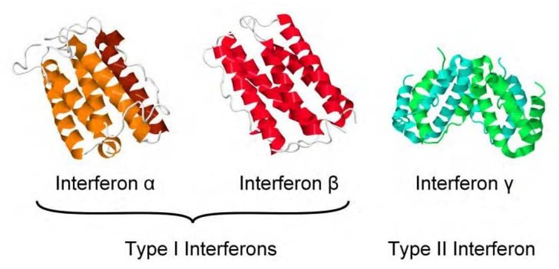
Interferons: Interferons (IFNs) are a family of antiviral and immunomodulatory signaling proteins produced by host cells to fight pathogens like viruses, bacteria, and tumors

(IFNs) are a family of antiviral and immunomodulatory signaling proteins produced by host cells to fight pathogens like viruses, bacteria, and tumors.

As cytokines, they alert neighboring cells to activate defenses, inhibit viral replication, and regulate immune responses.

Common uses include treating hepatitis B and C, multiple sclerosis, and certain cancers like melanoma and lymphoma.

For more information click on the link below: sciencenewshighlights ScienceMission.

Why you can’t tie knots in four dimensions

We all know we live in three-dimensional space. But what does it mean when people talk about four dimensions? Is it just a bigger kind of space? Is it “space-time,” the popular idea which emerged from Einstein’s theory of relativity?

If you have wondered what four dimensions really look like, you may have come across drawings of a “four-dimensional cube.” But our brains are wired to interpret drawings on flat paper as two-or at most three-dimensional, not four-dimensional.

The almost insurmountable difficulty of visualizing the fourth dimension has inspired mathematicians, physicists, writers and even some artists for centuries. But even if we can’t quite imagine it, we can understand it.

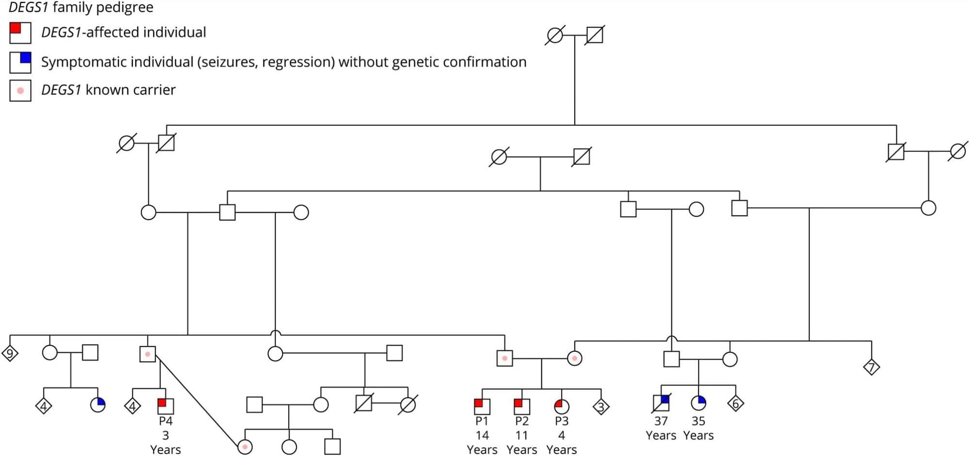
DEGS1-Related Hypomyelinating LeukodystrophyFour Individuals From Same Family and Review of Literature

Degs1-related hypomyelinating leukodystrophy: four individuals from same family and review of literature.


ObjectivesDEGS1-related leukodystrophy has been described in 33 individuals to date with varying degrees of neurologic symptoms and atrophy/hypomyelination on MRI brain. We describe a family of 4 individuals with DEGS1-related leukodystrophy with phenotypic heterogeneity both within the family and in previously reported individuals.

Management of Inherited CNS Small Vessel Diseases: The CADASIL Example: A Scientific Statement From the American Heart Association

Lacunar infarcts and vascular dementia are important phenotypic characteristics of cerebral autosomal dominant arteriopathy with subcortical infarcts and leukoencephalopathy, the most common inherited cerebral small vessel disease. Individuals with the disease show variability in the nature and onset of symptoms and rates of progression, which are only partially explained by differences in pathogenic mutations in the NOTCH3 gene. Recognizing the disease early in its course and securing a molecular diagnosis are important clinical goals, despite the lack of proven disease-modifying treatments.

Frequently distracted? Your brain rhythms may be to blame

Scientists may have new answers to why pop-ups or notifications grab our attention. Turns out our attention is on a cycle, shifting seven to 10 times per second. This rhythmic occurrence may be crucial for survival, as it prevents us from becoming overly focused on one thing in our environment. It could help us to see a car backing up in a parking lot while we search for where we parked, or to duck to avoid a low-hanging tree branch on a walk while watching a kid ride a bike.

However, these windows that shift our attention could also make us more susceptible to distractions, especially in modern times. As we live in a world surrounded by screens, digital alerts, and other visual stimuli, these frequent and innate windows for shifting attention may make it easier to be pulled away from a task.

“For our ancestors who had to continue to monitor the environment for predators while foraging for food, this was a beneficial trait,” said Ian Fiebelkorn, Ph.D., assistant professor of Neuroscience at the Del Monte Institute for Neuroscience at the University of Rochester and senior author of a study out in the journal PLOS Biology. “But in our modern environment, with laptops open in front of us and a smartphone nearby, rhythmically occurring windows for beneficial attentional shifts might also work against us. That is, rhythmically occurring windows for attentional shifts are also associated with increased susceptibility to distracting information.”

/* */