Toggle light / dark theme

New Research Reveals That a Single Session of Exercise Can Supercharge Your Brain for the Next 24 Hours

Exercise improves cognitive performance for over 24 hours, especially when paired with good sleep. A study of older adults links physical activity and deep sleep to better memory, highlighting the importance of an active lifestyle for brain health.

Exercise provides a short-term boost to brain function that can last throughout the next day, according to a new study by researchers at University College London (UCL).

Earlier research conducted in controlled laboratory settings revealed that cognitive performance improves in the hours following exercise. However, the duration of these benefits remained unclear.

Human thought crawls at 10 bits per second, Caltech study finds

Researchers at the California Institute of Technology have unveiled a startling revelation about the human mind: our thoughts move at a mere 10 bits per second, a rate that pales in comparison to the staggering billion bits per second at which our sensory systems gather environmental data. This discovery, published in the journal Neuron, is challenging long-held assumptions about human cognition.

The research, conducted in the laboratory of Markus Meister, the Anne P. and Benjamin F. Biaggini Professor of Biological Sciences at Caltech, and spearheaded by graduate student Jieyu Zheng, applied information theory techniques on an extensive collection of scientific literature. By analyzing human behaviors such as reading, writing, video gaming, and Rubik’s Cube solving, the team calculated the 10 bits per second figure – a rate that Meister describes as “extremely low.”

To put this in perspective, a typical Wi-Fi connection processes about 50 million bits per second, making our thought processes seem glacial by comparison. This stark contrast raises a paradox that Meister and his team are eager to explore further: “What is the brain doing to filter all of this information?”

Microbes Have Memory: Surprising Parallels Between Simple Microrganisms and Sophisticated Neurons

Discovery draws surprising parallels between low-level organisms and sophisticated neurons; lays the groundwork for memory-capable biological systems.

Biologists studying collectives of bacteria, or “biofilms,” have discovered that these so-called simple organisms feature a robust capacity for memory.

Working in the laboratory of University of California San Diego Professor Gürol Süel, Chih-Yu Yang, Maja Bialecka-Fornal and their colleagues found that bacterial cells stimulated with light remembered the exposure hours after the initial stimulus. The researchers were able to manipulate the process so that memory patterns emerged.

ADULT NEUROGENESIS IN HUMANS: A Review of Basic Concepts, History, Current Research, and Clinical Implications

The evidence presented by Boldrini et al2 was considered resilient and convincing because the authors ensured that the samples were taken from healthy individuals using more biological parameters, such as angiogenesis and change in volume of DG, for a suitable comparison. They also employed unbiased stereology, which is the gold standard for counting number of neurons.2

The contrary results presented by the Sorrels study and Boldrini study highlight the existing ambiguity regarding the concept of neurogenesis in adult humans. Both studies employed reasonably similar immunohistological methods and included many of the same neurogenesis markers, yet contrasting results were observed. The study by Boldrini et al examined samples from humans aged 14 to 79 years, finding more than a thousand cells in each part of the DG, which was in stark contrast to what was observed by Sorrells et al, who found very few cells in the neurogenic niche of subjects in the same age range. Even if we consider that some discrepancies in numbers might arise due to the difference in counting methods (or subjective reasons), such marked and obvious disparity is perplexing. Sorrels et al justified their findings, noting the limitation that relying solely on the presence of markers might cause glial lineage cells to be identified as neuronal lineage cells. However, the authors stated that they used additional methods for confirmation, including transmission electron microscopy (TEM)-immunogold and in-situ hybridization. The relative paucity of any type of progenitor or immature cells, including glia in the neurogenic niche of DG in their study, remains unexplained.

Kempermann et al7 expressed skepticism regarding the negative findings of Sorrells et al, naming the postmortem interval, the lack of known status regarding neuropsychiatric disease or chronic ailment, and using patients with epilepsy as key factors of concern.7 Kempermann et al argued that, in severe epilepsy, destruction of the neurogenic niche is an explicit possibility, and that, in some cases, epilepsy could be the reason for the lack of neurogenesis.7 Additionally, Kempermann et al also criticized the use of 10% formalin in some of the samples due its potential to mask the expression of proteins.7 However, Sorrells et al clearly mentioned that they performed appropriate antigen retrieval for the selected sections.1 Kempermann et al7 suggested that dependence on the protein markers to denote neurogenesis could be an erroneous approach and that some of the markers, such as DCX, are known for fast degradation. Additionally, the presence of DCX-negative immature neurons and high inter-individual variation in expression of DCX in humans, which was also reflected in the data presented by Boldrini et al, are not unusual.7 The use of fluorescence markers also was presented as a caveat by Kempermann et al7 because it is prone to fade away and might give rise to false negative impression.

Stem cells head to the clinic: treatments for cancer, diabetes and Parkinson’s disease could soon be here

Andrew Cassy had spent his working life in a telecommunications research department until a diagnosis of Parkinson’s disease in 2010 pushed him into early retirement. Curious about his illness, which he came to think of as an engineering problem, he decided to volunteer for clinical trials.

“I had time, something of value that I could give to the process of understanding the disease and finding good treatments,” he says.

In 2024, he was accepted into a radical trial. That October, surgeons in Lund, Sweden, placed neurons that were derived from human embryonic stem (ES) cells into his brain. The hope is that they will eventually replace some of his damaged tissue.

The study is one of more than 100 clinical trials exploring the potential of stem cells to replace or supplement tissues in debilitating or life-threatening diseases, including cancer, diabetes, epilepsy, heart failure and some eye diseases. It’s a different approach from the unapproved therapies peddled by many shady clinics, which use types of stem cell that do not turn into new tissue.


More than 100 clinical trials put stem cells for regenerative medicine to the test. It’s a turning point for a field beset with ethical and political controversy.

Tiny, wireless antennas use light to monitor cellular communication

Monitoring electrical signals in biological systems helps scientists understand how cells communicate, which can aid in the diagnosis and treatment of conditions like arrhythmia and Alzheimer’s.

But devices that record electrical signals in cell cultures and other liquid environments often use wires to connect each electrode on the device to its respective amplifier. Because only so many wires can be connected to the device, this restricts the number of recording sites, limiting the information that can be collected from cells.

MIT researchers have now developed a biosensing technique that eliminates the need for wires. Instead, tiny, wireless antennas use light to detect minute electrical signals.

Clinical trial shows propranolol reduces tremors in Parkinson’s disease

The standard medication levodopa does not always work against tremors in Parkinson’s disease, especially in stressful situations. Propranolol, however, does work during stress, providing insight into the role of the stress system in tremors. MRI scans reveal that propranolol directly inhibits activity in the brain circuit that controls tremors. Doctors may consider this medication when levodopa is ineffective.

People with Parkinson’s disease report that worsen during stressful situations. “Tremors act as a sort of barometer for stress; you see this in all people with Parkinson’s,” says neurologist Rick Helmich from Radboud university medical center.

The commonly used drug levodopa usually helps with tremors, but it tends to be less effective during stress, when tremors are often at their worst. Helmich and his team wanted to investigate whether a medication targeting the stress system could help and how this effect of stress on tremors works in the brain. The work is published in the journal Annals of Neurology.

Long COVID: SARS-CoV-2 Spike Protein Accumulation Linked to Long-lasting Brain Effects

Researchers from Helmholtz Munich and Ludwig-Maximilians-Universität (LMU) have identified a mechanism that may explain the neurological symptoms of long COVID.

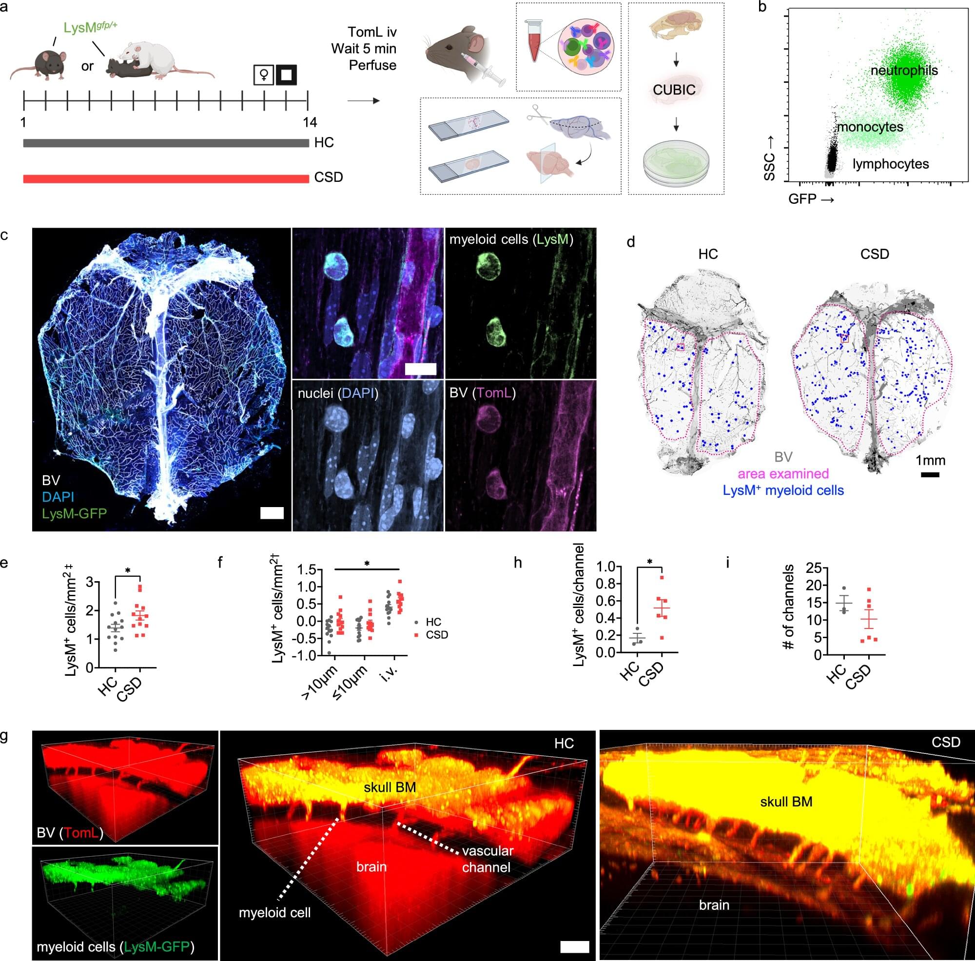
The study shows that the SARS-CoV-2 spike protein remains in the brain’s protective layers, the meninges, and the skull’s bone marrow for up to four years after infection. This persistent presence of the spike protein could trigger chronic inflammation in affected individuals and increase the risk of neurodegenerative diseases.

The team, led by Prof. Ali Ertürk, Director at the Institute for Intelligent Biotechnologies at Helmholtz Munich, also found that mRNA COVID-19 vaccines significantly reduce the accumulation of the spike protein in the brain. However, the persistence of spike protein after infection in the skull and meninges offers a target for new therapeutic strategies.