New research shows that older adults may compensate for age-related cognitive decline by enhancing activity in a specific brain region linked to attention—the locus coeruleus (LC).

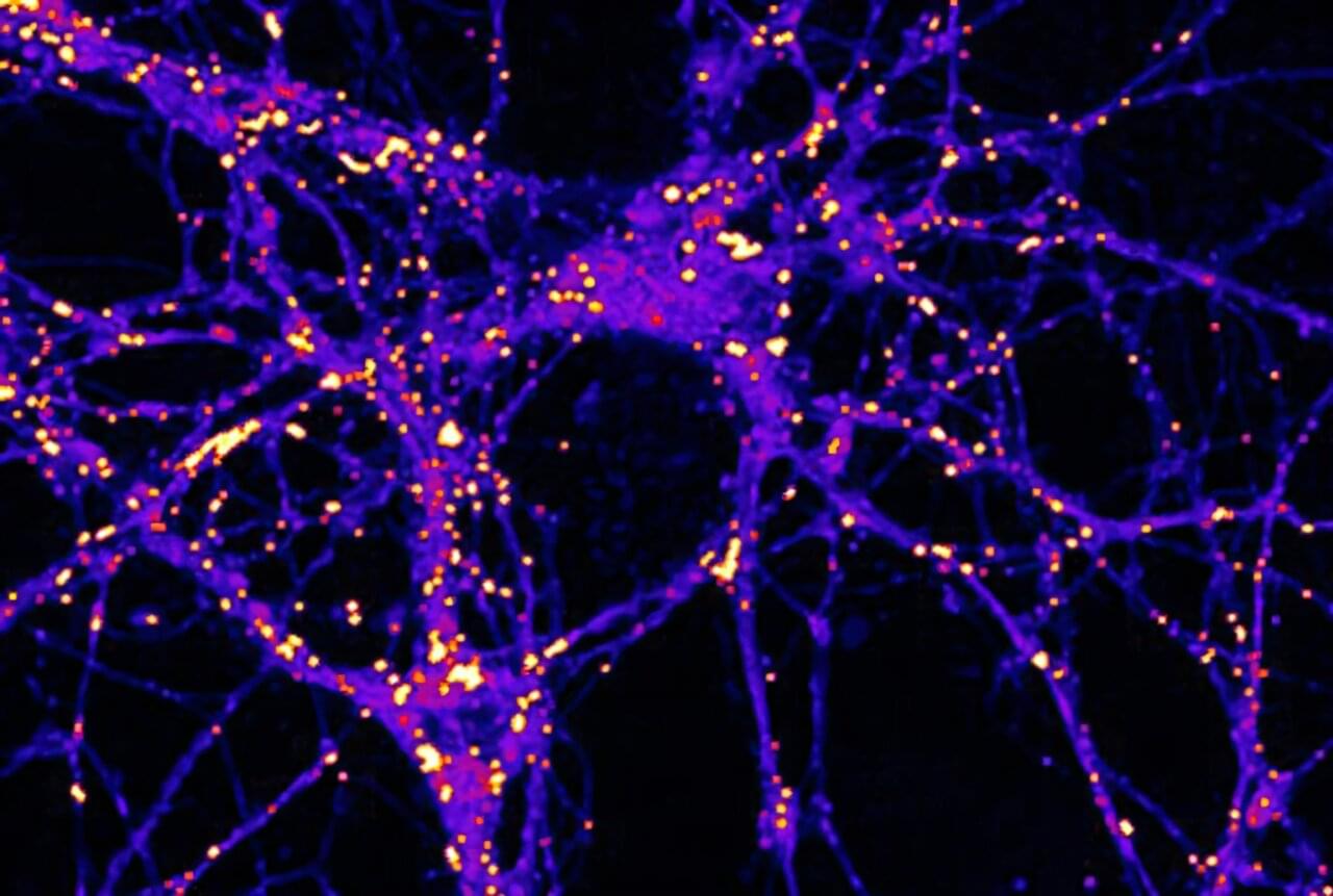
Cellular communication between neurons within our brain is complex and busy, much like a USPS mailroom.
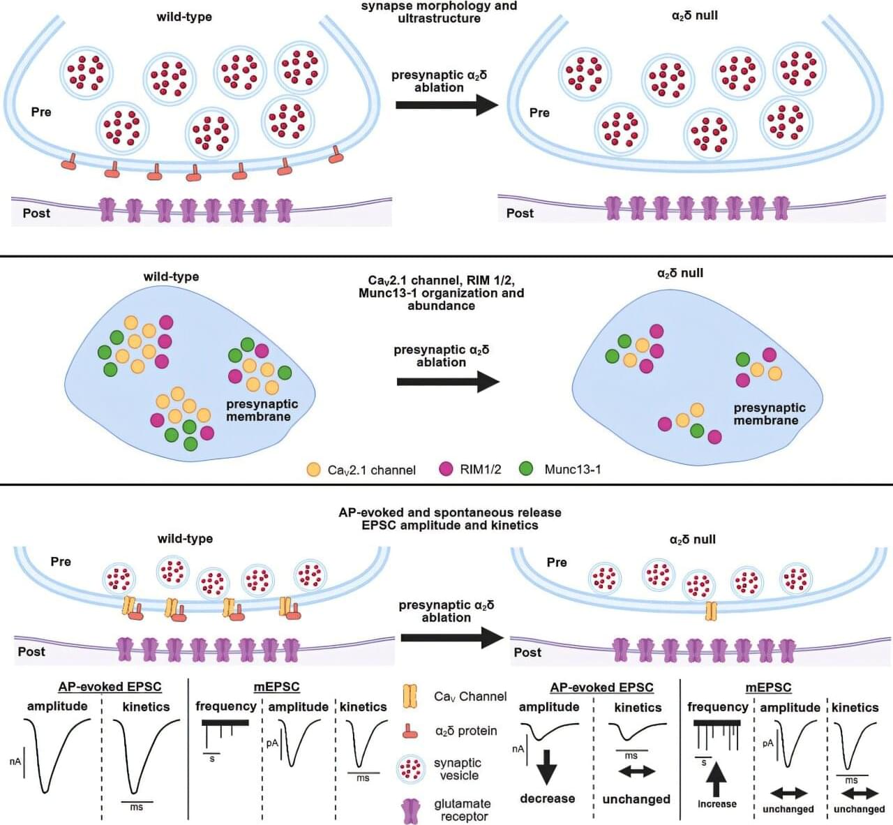
To keep things running smoothly, the brain uses specialized molecules, termed alpha-2-delta (α2δ) proteins, to coordinate the sending and receival of signals between nerve cells in the brain.
Genetic variations in these types of proteins can impact important brain messaging and function, resulting in chronic pain, autism spectrum disorders, epilepsy, migraines, and other conditions.
While glucose, or sugar, is a well-known fuel for the brain, Weill Cornell Medicine researchers have demonstrated that electrical activity in synapses—the junctions between neurons where communication occurs—can lead to the use of lipid or fat droplets as an energy source.
The study, published in Nature Metabolism, challenges “the long-standing dogma that the brain doesn’t burn fat,” said principal investigator Dr. Timothy A. Ryan, professor of biochemistry and of biochemistry in anesthesiology, and the Tri-Institutional Professor in the Department of Biochemistry at Weill Cornell Medicine.
The paper’s lead author, Dr. Mukesh Kumar, a postdoctoral associate in biochemistry at Weill Cornell Medicine who has been studying the cell biology of fat droplets, suggested that it makes sense that fat may play a role as an energy source in the brain like it does with other metabolically demanding tissues, such as muscle.
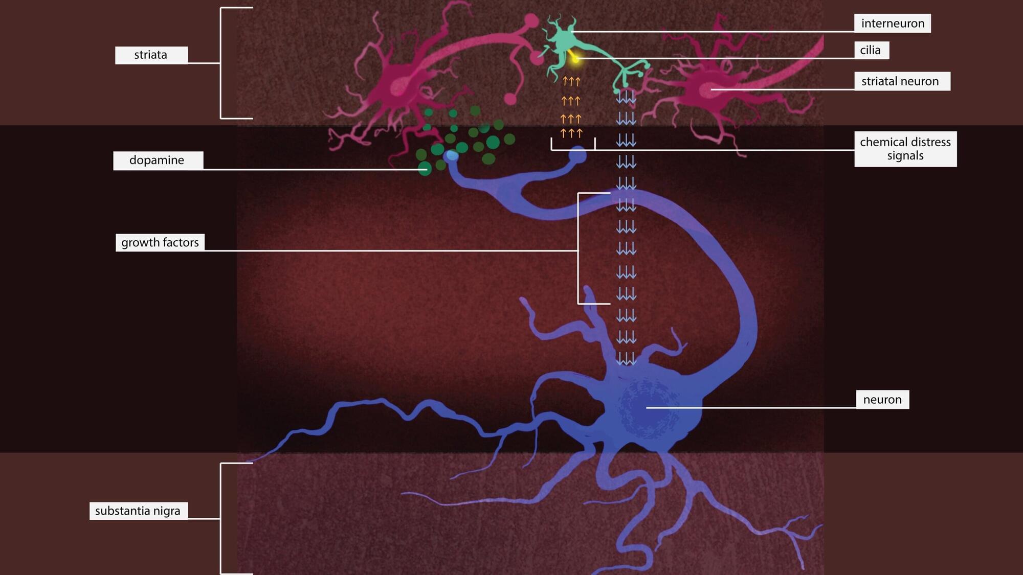
Putting the brakes on an enzyme might rescue neurons that are dying due to a type of Parkinson’s disease that’s caused by a single genetic mutation, according to a new Stanford Medicine-led study conducted in mice.
The study has been published in Science Signaling.
The genetic mutation causes an enzyme called leucine-rich repeat kinase 2, or LRRK2, to be overactive. Too much LRRK2 enzyme activity changes the structure of brain cells in a way that disrupts crucial communication between neurons that make the neurotransmitter dopamine and cells in the striatum, a region deep in the brain that is part of the dopamine system and is involved in movement, motivation and decision-making.

Leafcutter ants live in highly organized colonies where every ant has a job, and now researchers can flip those jobs like a switch. By manipulating just two neuropeptides, scientists can turn defenders into nurses or gardeners into leaf harvesters. These same molecular signals echo in naked mole-rats, revealing a deep evolutionary link in how complex societies function, even across species. The study also teases out a possible connection to insulin and longevity, hinting at new frontiers in understanding human behavior and lifespan.
To study what happened in the brain during this task, the researchers used functional magnetic resonance imaging, which measures blood flow as a proxy for neural activity. They scanned the brains of 11 participants while they performed the memory task over multiple sessions. By applying a complex decoding model to the imaging data, the researchers were able to estimate not only what participants were remembering but also how uncertain they were about each memory. The model treated neural activity as a probabilistic code, where stronger or more focused patterns of activity reflected more confident memory representations.
The results showed that neural signals in the visual cortex—the area of the brain involved in processing visual information—were more intense for the high-priority memory items. These stronger signals translated to smaller memory errors and greater confidence. On average, participants remembered the high-priority items more accurately and responded more quickly when asked to recall them. Their eye movements were closer to the correct location, and they took less time to respond. These behavioral improvements matched the patterns observed in the brain data.
The study also found that the magnitude of neural activity in the frontal cortex predicted how well participants could distinguish between high-and low-priority memories. This suggests that the frontal cortex plays a regulatory role, sending signals that adjust the strength of memory representations in visual areas depending on how important each item is. In other words, the frontal brain regions help direct the mental spotlight, increasing the “volume” of the memories that matter most.
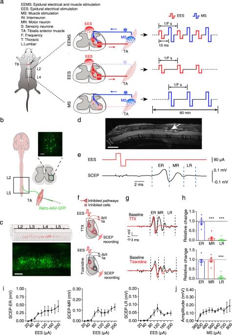
Electrical signals with characteristic parameters for reconstructing neural circuits remain incompletely understood, limiting the therapeutic potential of electrical neuromodulation techniques. Here, the authors demonstrate that dual electrical stimulation at 10–20 Hz rebuilds the spinal sensorimotor neural circuit after spinal cord injury, indicating the characteristic signals of circuit remodeling.
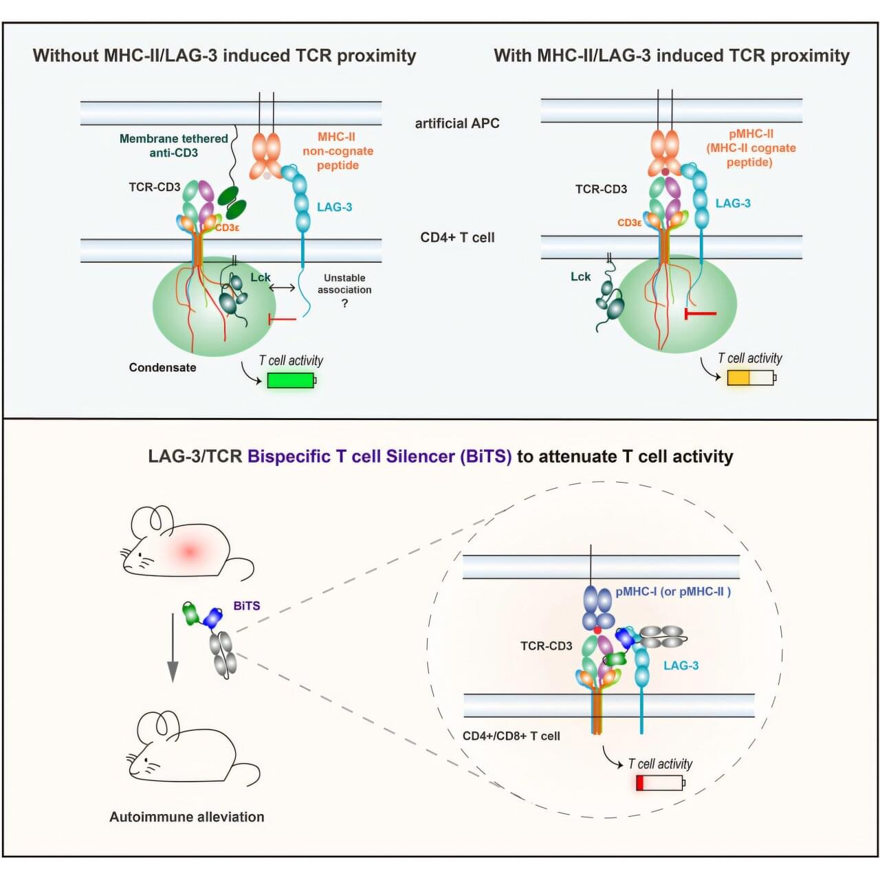
An engineered protein turns off the kind of immune cells most likely to damage tissue as part of type-1 diabetes, hepatitis, multiple sclerosis, shows a new study in mice.
In these autoimmune diseases, T cells mistakenly target the body’s own tissues instead of invading viruses or bacteria as they would during normal immune responses. Treatments focused on T cells have been elusive because blocking their action broadly weakens the immune system and creates risk for infections and cancer.
Published online June 30 in the journal Cell, the study revealed that holding closely together two protein groups (signaling complexes) on T cells, including one found more often on T cells involved in autoimmune disease, shuts down those T cells in a limited way.

A team of researchers at NYU Abu Dhabi has uncovered a key mechanism that helps shape how our brains are wired, and what can happen when that process is disrupted.
In a new study published in Cell Reports, the RNA-MIND Lab at NYU Abu Dhabi, led by Professor of Biology Dan Ohtan Wang, with Research Associate Belal Shohayeb, reveals how a small molecular mark on messenger RNA, called m6A methylation, regulates the production of essential proteins inside growing neurons. This process plays a critical role in the development of axons, the long extensions that neurons use to connect and communicate with each other.
The study shows that this molecular mark controls the production of a protein called adenomatous polyposis coli (APC), which helps organize the internal structure of nerve cells and is needed to locally produce β-actin, a key building block of the cytoskeleton to support axon growth. Importantly, the team also found that genetic mutations linked to autism and schizophrenia can interfere with this process, potentially affecting how the brain develops.