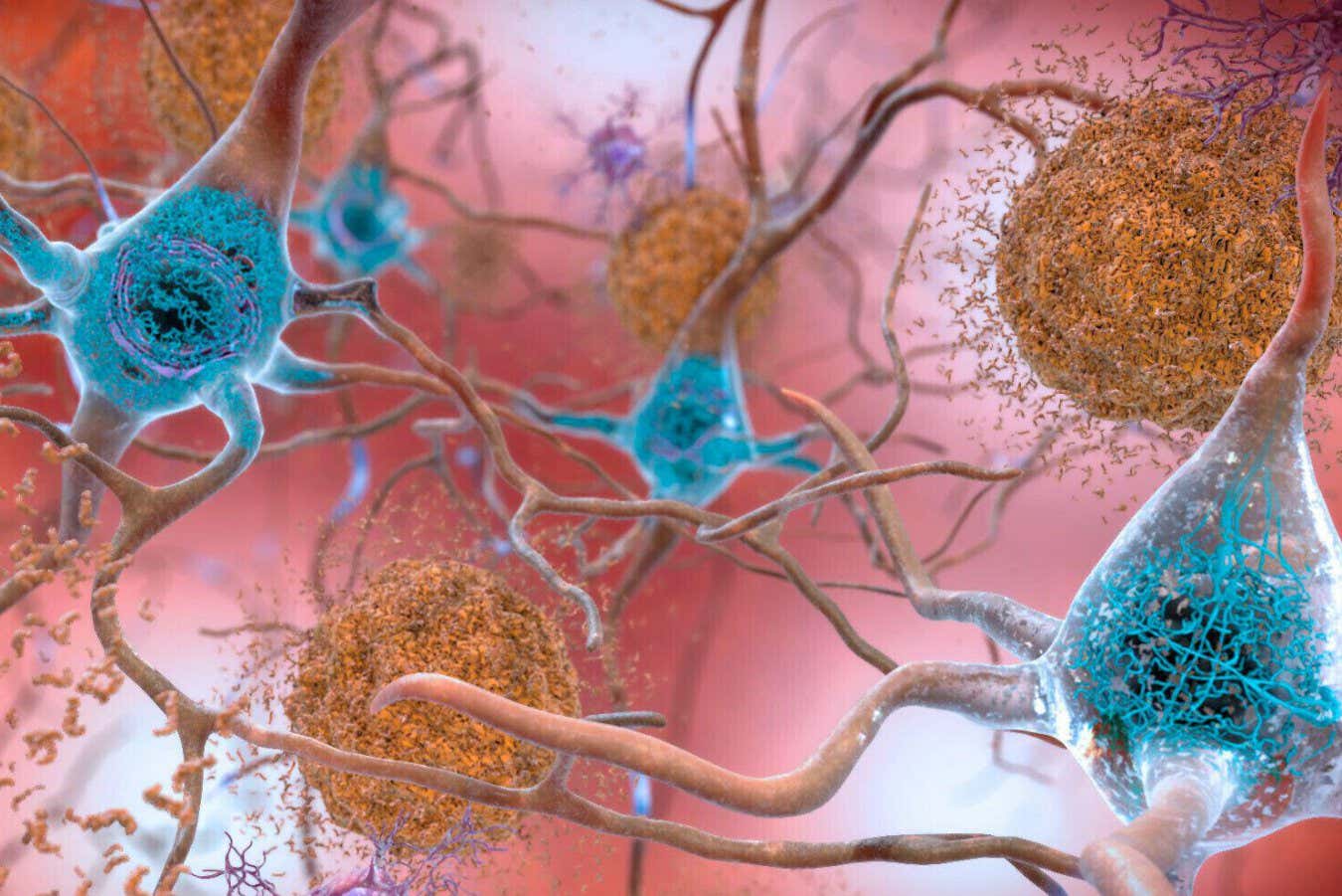
Toggle light / dark theme

APOE ε4 variant reveals hidden risk factors beyond Alzheimer’s

Further analysis for immune-specific processes revealed APOE ε4 enrichment in various infection-related pathways, including herpes, influenza A, hepatitis, measles, and Epstein-Barr virus (EBV). Significant enrichment was also observed for B-cell, T-cell, and inflammatory signaling cascades. Next, immune cell subtype enrichment analysis revealed the most APOE ε4 enrichment in intermediate and non-classical monocytes among innate immune cells.

Among adaptive immune cells, memory cluster of differentiation 8 (CD8) T cells, regulatory T (Treg) cells, and memory CD4 T cells were the most enriched. Besides, γδ T cells and natural killer (NK) cells showed APOE ε4 enrichment. In the liver, a cell-type-specific enrichment analysis revealed the most APOE ε4 enrichment in Kupffer cells and hepatocytes.

Next, the researchers examined whether APOE ε4 CSF proteome changes were reflected in the plasma and used the GNPC dataset for plasma proteome profiling of AD, PDD, FTD, PD, ALS, and non-impaired controls. Fifty-eight plasma proteins associated with the APOE genotype were identified in non-impaired controls. CART modeling revealed that these 58 proteins could strongly differentiate between APOE ε4 carriers and non-carriers across neurodegenerative diseases, and this signature was found to be consistent across different sexes and racial groups.

Psychopathic traits linked to distinct brain networks in new neuroscience research

Psychopathy is often associated with impulsivity, aggression, and antisocial behavior. While past studies have focused heavily on how different brain regions function in people with psychopathic traits, less is known about how these regions are structurally connected. Structural connectivity refers to the physical links between brain areas—similar to the brain’s wiring system. The researchers aimed to go beyond earlier work that focused only on specific brain circuits and instead look across the entire brain to identify any structural patterns linked to psychopathy.

The researchers were especially interested in understanding whether structural differences in the brain might explain the relationship between psychopathic traits and externalizing behaviors. Previous models have suggested two possible brain-based explanations for these behaviors. One theory emphasizes problems in how people process emotional threats, while another highlights difficulties in attention control. Both theories have some support, but no study had comprehensively examined how structural brain networks might connect psychopathy with real-world behavioral problems.

The research team analyzed data from 82 young adults who participated in the Leipzig Mind-Brain-Body study. All participants were screened to rule out medical or psychological conditions that might affect the results. Psychopathic traits were assessed using a questionnaire designed to capture both interpersonal-affective characteristics (like manipulation and lack of empathy) and behavioral traits (like impulsivity and rule-breaking). Externalizing behaviors were also measured with a separate questionnaire that included items on aggression, defiance, and similar tendencies.

Each participant underwent high-resolution brain imaging using diffusion MRI, a technique that maps the white matter tracts—essentially the brain’s wiring—connecting different regions. The researchers used a method called connectome-based predictive modeling, which relies on machine learning to identify patterns in the brain’s structural connectivity that relate to individual differences in behavior.

This method allowed them to identify two kinds of networks: positive networks, where stronger connections were linked to higher psychopathy scores, and negative networks, where weaker connections were related to those same scores. They also tested whether specific connections within these networks helped explain the relationship between psychopathic traits and externalizing behaviors.

The results showed that psychopathic traits were significantly associated with both stronger and weaker connections in different parts of the brain. The positive network—made up of connections that increased with psychopathy—was better at predicting psychopathic traits than the negative network alone. But when both networks were combined, the prediction became even more accurate.

Many of the connections in the positive network were located within the brain’s frontal and parietal lobes, which are involved in decision-making, emotional processing, and attention. These connections included pathways like the uncinate fasciculus, which links the frontal cortex with areas involved in emotion, and the arcuate fasciculus, which supports language and auditory processing. Other connections involved the cingulum bundle, associated with emotional regulation and social behavior, and the posterior corticostriatal pathway, which plays a role in reward processing and learning.

‘Universal cancer vaccine’ trains the immune system to kill any tumor

Following on from their breakthrough human trial that successfully reprogrammed the immune system to overpower glioblastoma, an aggressive brain tumor, the same scientists have now further developed the mRNA vaccine to fight not one but any cancer. It has the potential to do away with chemotherapy, surgery and radiation treatment.

University of Florida (UF) scientists have developed an experimental vaccine that dramatically boosts the immune system’s ability to fight tumors – even without targeting a specific cancer type. This “general purpose” mRNA jab works in a similar way to a Covid-19 vaccine but with a different target; it instructs the body’s immune cells to rally and hit any kind of tumor in the same way they would attack a viral spike protein.

“This paper describes a very unexpected and exciting observation: that even a vaccine not specific to any particular tumor or virus – so long as it is an mRNA vaccine – could lead to tumor-specific effects,” said Elias Sayour, a pediatric oncologist and principal investigator at the RNA Engineering Laboratory at UF. “This finding is a proof of concept that these vaccines potentially could be commercialized as universal cancer vaccines to sensitize the immune system against a patient’s individual tumor.”

Wellesley team’s new research on anesthesia unlocks important clues about the nature of consciousness

For decades, one of the most fundamental and vexing questions in neuroscience has been: what is the physical basis of consciousness in the brain? Most researchers favor classical models, based on classical physics, while a minority have argued that consciousness must be quantum in nature, and that its brain basis is a collective quantum vibration of “microtubule” proteins inside neurons.

New research by Wellesley College professor Mike Wiest and a group of Wellesley College undergraduate students has yielded important experimental results relevant to this debate, by examining how anesthesia affects the brain. Wiest and his research team found that when they gave rats a drug that binds to microtubules, it took the rats significantly longer to fall unconscious under an anesthetic gas. The research team’s microtubule-binding drug interfered with the anesthetic action, thus supporting the idea that the anesthetic acts on microtubules to cause unconsciousness.

“Since we don’t know of another (i.e,. classical) way that anesthetic binding to microtubules would generally reduce brain activity and cause unconsciousness,” Wiest says, “this finding supports the quantum model of consciousness.”

The Role of Innovation Technology in the Rehabilitation of Patients Affected by Huntington’s Disease: A Scoping Review

Huntington’s disease is an autosomal dominant neurodegenerative disease caused by the repetition of cytosine, adenine, and guanine trinucleotides on the short arm of chromosome 4p16.3 within the Huntingtin gene. In this study, we aim to examine and map the existing evidence on the use of innovations in the rehabilitation of Huntington’s disease. A scoping review was conducted on innovative rehabilitative treatments performed on patients with Huntington’s disease. A search was performed on PubMed, Embase, Web of Science, and Cochrane databases to screen references of included studies and review articles for additional citations. Of an initial 1,117 articles, only 20 met the search criteria. These findings showed that available evidence is still limited and that studies generally had small sample sizes and a high risk of bias.

Cognitive Stimulants: from Caffeine to Cannabinoids — Current and Future Perspectives

Habitual coffee consumers justify their life choices by arguing that they become more alert and increase motor and cognitive performance and efficiency; however, these subjective impressions still do not have a neurobiological correlation. Using functional connectivity approaches to study resting-state fMRI data in a group of habitual coffee drinkers, we herein show that coffee consumption decreased connectivity of the posterior default mode network (DMN) and between the somatosensory/motor networks and the prefrontal cortex, while the connectivity in nodes of the higher visual and the right executive control network (RECN) is increased after drinking coffee; data also show that caffeine intake only replicated the impact of coffee on the posterior DMN, thus disentangling the neurochemical effects of caffeine from the experience of having a coffee.

There is a common expectation, namely among habitual coffee drinkers, that coffee increases alertness and psychomotor functioning. For these reasons, many individuals keep drinking coffee to counteract fatigue, stay alert, increase cognitive performance, and increase work efficiency (Smith, 2002). Coffee beverages are constituted of numerous compounds known to affect human behavior, among which are caffeine and chlorogenic acids (Sadiq Butt et al., 2011). From the neurobiological perspective, both caffeine and chlorogenic acids have well-documented psychoactive actions, whereas caffeine is mostly an antagonist of the main adenosine receptors in the brain—A1 and A2A receptors, leading to the disinhibition of excitatory neurotransmitter release and enhancement of dopamine transmission via D2 receptors (Fredholm et al., 2005) to sharpen brain metabolism and bolster memory performance (Paiva et al.

/* */