Toggle light / dark theme

Lab-grown pineal gland organoids produce melatonin, offering a new sleep model

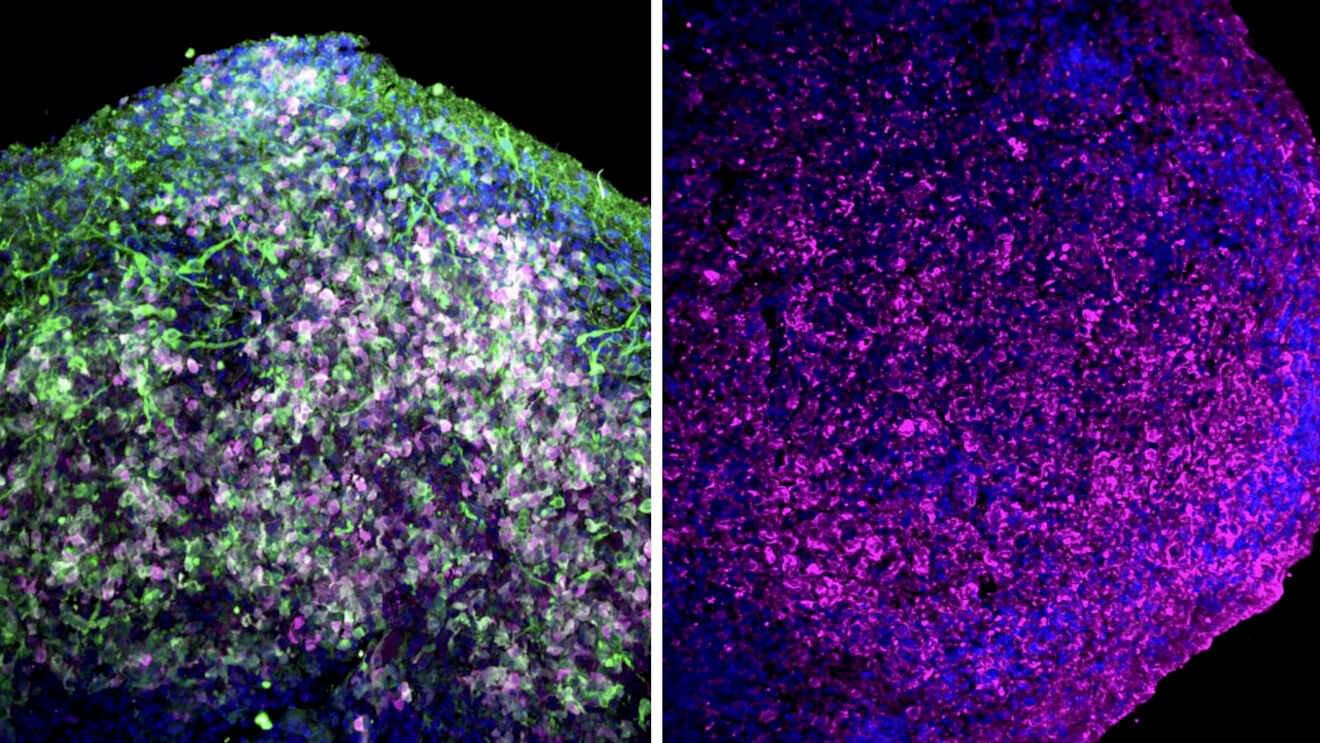
Organoids are miniature, simplified versions of an organ. Over the past two decades, scientists have developed them for the gut, lung, liver, mammary gland, brain, and more. Now, researchers at Yale School of Medicine (YSM) have organoid-ized the pineal gland, a small structure in the brain that regulates sleep patterns through its production of the hormone melatonin.

In a study published in Cell Stem Cell, the researchers demonstrate how pineal gland organoids can be used to study sleep dysfunction in conditions like Angelman syndrome, autism, and depression.

“In a number of neuropsychiatric conditions, severe sleep problems are a major symptom,” says In-Hyun Park, Ph.D., associate professor of genetics at YSM and senior author of the study. “With pineal gland organoids, we may be able to uncover the causes of those sleep disturbances and possibly identify treatments.”

Sound-sensing hair bundles in our ears act as tiny thermodynamic machines

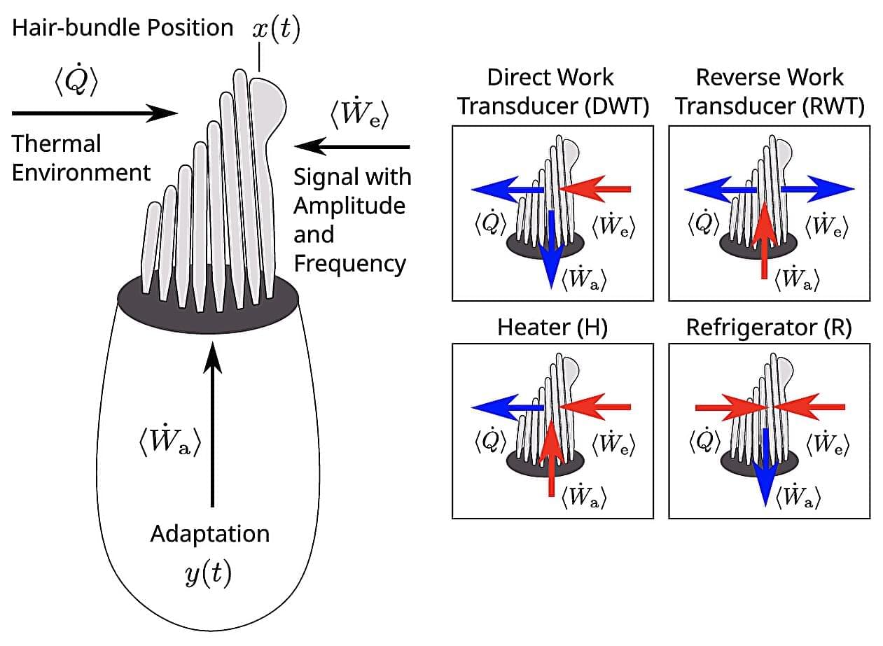
The hair cells lining the inner ear are among the most sophisticated structures in the human body: capable of detecting sounds as faint as a whisper, while helping to maintain our sense of balance. Through new models detailed in PRX Life, a team led by Roman Belousov at the European Molecular Biology Laboratory has revealed for the first time how oscillating bundles attached to these cells operate in different thermodynamic regimes—offering a new framework for understanding how our hearing works at a fundamental level.

Within the inner ear, each hair cell hosts a hair “bundle”: a cluster of tiny, bristle-like projections that vibrate in response to incoming sound waves. The mechanical energy from these oscillations is then converted into electrical signals which travel to the brain. Rather than being passive receivers, these bundles actively oscillate —driven by molecular motors within the cell that allow them to amplify faint signals and tune in to specific frequencies.

But despite decades of study, researchers are still unclear on the connection between this active oscillation and the hair bundle’s response to external sound. Existing models tended to treat bundles as if they were moving spontaneously, without accounting for what happens when they actually interact with sound.

Mapping mutations at scale in a single gene reveals new neurodevelopmental condition

The ability of different genetic variants—changes to one or more building blocks of DNA—to cause disease, and to what extent, has historically been opaque. Geneticist and Crick group leader Greg Findlay has pioneered a new method in the hope of changing this. Called “saturation genome editing,” the new technique involves mapping every single variant in a given gene to work out what it does and pinpoint which changes are responsible for specific disorders.

While Greg was refining these experiments, Nicky Whiffin, associate professor at the University of Oxford, had identified that mutations in a tiny gene were behind a rare inherited neurodevelopmental disorder, known as ReNU syndrome, which impacts brain function, development and motor skills. Children develop this syndrome if a single copy of the RNU4-2 gene is mutated in a specific way.

Nicky initially found that several distinct mutations in a critical region of the gene caused the condition, and she was keen to understand if some of these genetic variants led to more severe disease.

APOE4 Increases Neurons’ Excitability Before Symptoms Appear

The pro-Alzheimer’s allele APOE4 makes hippocampal neurons in mice smaller and hyperexcitable. This effect, which resembles epilepsy and accelerated aging, can be mitigated by manipulating a neuronal protein [1].

Before symptoms arise

Alzheimer’s disease begins long before symptoms appear, building silently for decades. The single strongest genetic risk factor for the common, late-onset form of Alzheimer’s is the ε4 variant of the apolipoprotein (APOE) gene, APOE4. Carrying a single copy of this variant (being heterozygous) roughly triples your Alzheimer’s risk; having two copies increases it about 12-fold.

How an Alzheimer’s Risk Gene Disrupts Brain Circuits Long Before Memory Loss

Researchers at the Gladstone Institute have uncovered the molecular mechanism by which APOE4 — the most significant genetic risk factor for Alzheimer’s disease, present in roughly a quarter of the population — begins damaging neural circuits well before any cognitive symptoms emerge. Studying young mice carrying the APOE4 variant, the team found that the gene triggers overproduction of the protein Nell2, which causes neurons to shrink and become hyperactive. Crucially, the degree of early neuronal hyperactivity predicted the severity of memory impairment later in life, even in animals that still showed normal learning and memory at the time of measurement. Strikingly, targeting Nell2 therapeutically was able to reverse these changes even in adult animals, demonstrating that the neurodegeneration is not irreversible and that a window for intervention may exist even after the disease process has begun. The team is currently continuing preclinical testing of this therapeutic strategy.


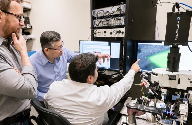
New findings on the APOE4 gene variant point to a potential therapeutic target for Alzheimer’s disease. From left to right, Gladstone scientists Misha Zilberter, Yadong Huang, and Dennis Tabuena examine findings from their research, which is published in the journal Nature Aging.

For the millions of people who carry the gene APOE4, the strongest known genetic risk factor for Alzheimer’s disease, their brain activity may begin changing long before any memory problems appear. Now, researchers at Gladstone Institutes have uncovered a precise chain of molecular events behind those early changes and identified a potential way to reverse them.

Published in the journal Nature Aging, their new study in mouse models reveals how APOE4 triggers increased production of the protein Nell2, which makes neurons shrink and become hyperactive. The more hyperactive the neurons were in early life, the more severe were the memory problems the mice developed later in life.

Frequency of AQP4 and MOG Antibodies in Patients With Optic Neuritis Fulfilling Minimal New Multiple Sclerosis MRI Criteria

How pancreatic cancer survives ferroptosis?

Pancreatic ductal adenocarcinoma (PDAC) tumors harboring KRAS mutations exhibit relative resistance to iron-dependent form of cell death, ferroptosis, compared with other tumor types but the mechanisms remain unclear.

The researchers reveal that hypoxia and pancreatic tumor interstitial fluid cooperate to suppress ferroptosis in pancreatic cancer through HIF-2 activity.

HIF-2 enables tumor survival by regulating glutathione metabolism through upregulating the expression of both components of the system Xc− cystine transporter and transsulfuration pathway enzymes CBS and CTH to increase intracellular cysteine levels.

HIF-2 also induces the Parkin mitophagy factor and suppresses mitochondrial function and reactive oxygen species (ROS) generation and thus survives metabolically hostile environments, defining a tissue-specific role in pancreatic ductal adenocarcinoma. sciencenewshighlights ScienceMission https://sciencemission.com/HIF-2-and-PDAC


Hubbi et al. reveal that hypoxia and pancreatic tumor interstitial fluid cooperate to suppress ferroptosis in pancreatic cancer through HIF-2 activity. By transcriptionally regulating glutathione metabolism and mitochondrial function, HIF-2 enables tumor survival in metabolically hostile environments, defining a tissue-specific role in pancreatic ductal adenocarcinoma.

Optogenetics, Biohybrid Implants And The Future Of Brain-Computer Interfaces | Dr. Alan Mardinly

Optogenetics, Biohybrid Implants And The Future Of Brain-Computer Interfaces — Dr. Alan Mardinly Ph.D. — CSO & Co-Founder, Science


What if we could restore vision, communicate directly with the brain, and even extend human life—not with machines alone, but with living, engineered biology?

Dr. Alan Mardinly, Ph.D. is the Chief Scientific Officer and Co-Founder of Science Corp. (https://science.xyz/), a neurotechnology company developing next-generation brain interfaces and biohybrid neural implants aimed at restoring human function.

Dr. Mardinly leads the company’s biohybrid program, focused on combining genetically engineered cells with advanced optical hardware to create optogenetic therapies for vision restoration and new types of brain-machine interfaces.

Dr. Mardinly has spent more than 15 years working at the intersection of neuroscience, genetics, and neural engineering.

Predicting Outcome After Newborn Stroke: A Lesion Network Mapping Study Leveraging Large-Scale Data

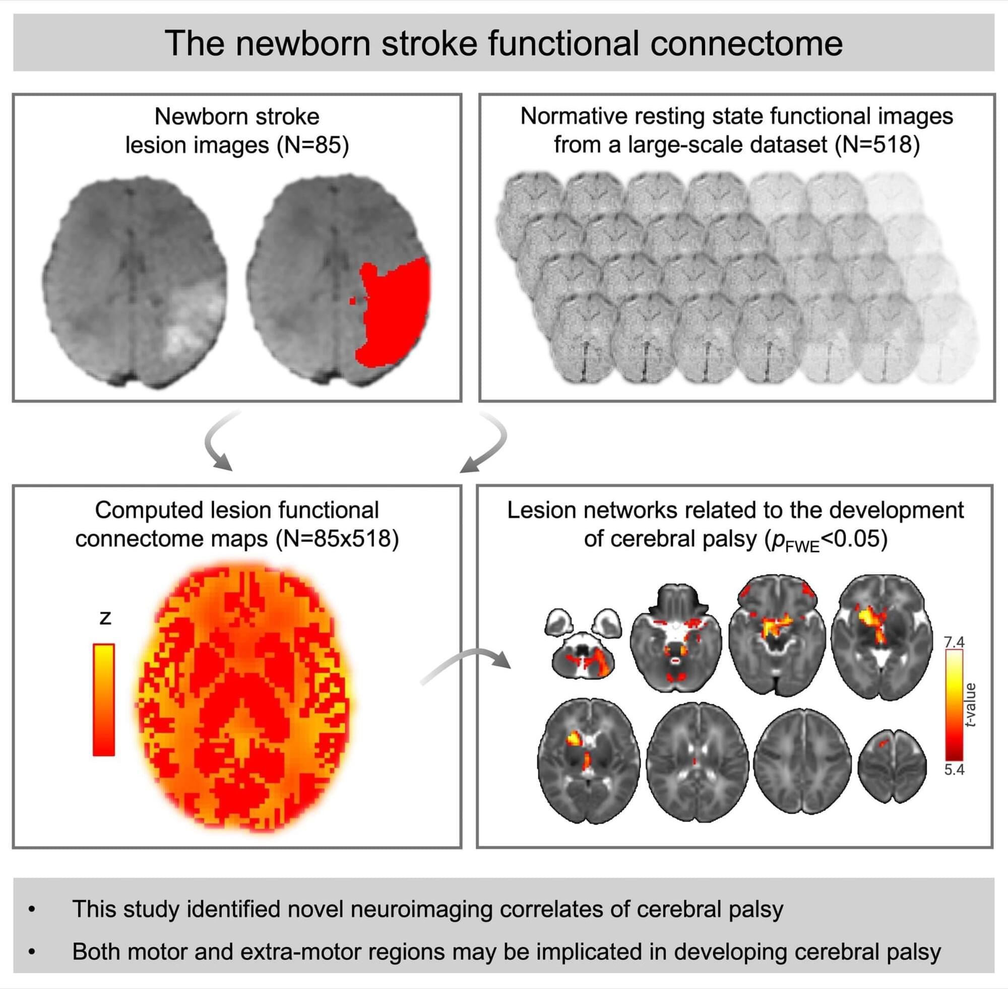
The development of cerebral palsy after neonatal stroke may be associated with disruptions of broad functional networks involving motor and extramotor regions as opposed to isolated lesions of the motor tracts.


Prior studies have established that resting-state networks are already present in newborns as early as term-equivalent age19 and that motor outcomes in healthy populations and other clinical populations are related to both primary motor networks and other broader motor networks.40,41 Our observation that the development of cerebral palsy correlated with both motor and extramotor regions lines up with these findings in other populations, and may indicate more widespread network vulnerabilities after stroke in neonates compared with adults.21 While it is known that disruption to primary motor regions is related to the development of cerebral palsy after NAIS,5,8 the extramotor regions (the frontal and temporal regions identified in the current study) may be more vulnerable to network-level disruptions in neonatal stroke due to the known relative immaturity of these regions during the neonatal period and their prolonged maturation to support higher-order functions.42,43 Particular vulnerability of these temporal and frontal regions has been demonstrated in other newborn, very preterm populations at risk of brain insult and injury.43

There are limitations to this study. Lesion network mapping has frequently been performed in adults and is more clinically feasible than directly acquiring resting-state fMRI scans in patients with NAIS; however, lesion network mapping may be seen as a less direct method for assessing brain connectivity.13,21,22 Functional connectivity has been well characterized in adults and neonates,1,16–19 but future studies will need to examine structural connectivity between regions via white matter fiber pathways using diffusion MRI scans to determine whether the functional changes identified correspond to structural changes.44 Such diffusion MRI investigations will be important in the future to investigate the roles (as part of global structural networks) of white matter regions and tracts such as the posterior limb of the internal capsule, which has previously been shown to be a strong predictor of motor outcome using individual region-based analyses.5–12 Given the paucity of prior research in this area, we aimed to establish that there is a relationship between the lesion functional connectome and cerebral palsy, but future work will also need to investigate the relative contributions of different measures (such as lesion volume, lesion location, and lesion functional and structural connectivity) to the development of cerebral palsy. Different measures may be more important for different brain regions (eg, lesions located in the primary motor regions are well-known to be related to cerebral palsy,5,8 whereas this study shows that functional connectivity of lesions to other subcortical and temporal and frontal cortical regions are related to cerebral palsy), meaning a combined approach considering multiple measures and regions may improve prediction of cerebral palsy in future work. Given that this study utilized clinically indicated MRIs from multiple sites for stroke participants, uniform scanner (eg, field strength) and sequence (eg, resolution) settings could not be used. The lesion network mapping approach does not necessarily require uniform scanning, making this approach more accessible. However, we acknowledge that variations in scanning parameters could influence some analysis steps, such as lesion segmentation and mapping to the template. To account for this, all analysis steps, including lesion segmentation and registration, were performed by experienced pediatric neurologists and image scientists, and outputs were extensively visually checked to ensure any poor-quality data were excluded.

This lesion network mapping approach identified correlations between lesions and the rest of the gray matter, which could include the equivalent lesion regions themselves (self-correlations, which are typically likely to be high). If self-correlations were very high in both groups (cerebral palsy and noncerebral palsy), then no significant group differences may be identified, which we think explains why the primary motor regions did not appear in the current findings (as the primary motor regions are key lesion regions affected by NAIS).5 The correlations identified between lesions and gray matter regions were positive (rather than negative correlations), which could reflect increased activity to support motor function (as opposed to decreased activity of irrelevant processes), as seen during task performance, but even intrinsically occurring at rest.45 Positive correlations are also, in general, stronger and less variable than negative correlations.46 We identified significant regions in both the left and right brain hemispheres; however, future work to specifically test whether there was a difference in the findings between hemispheres would be worthwhile (ie, to test whether results were greater in magnitude or spatial extent in the left or right hemisphere, which could be related to asymmetry in the lesions or connectome or both). With lesion network mapping, it was possible to infer that the regional networks identified correlated with cerebral palsy, but we cannot necessarily extrapolate on the relative importance of the individual regions of the network in cerebral palsy based on this method alone.13
/* */